PHTY101 Lecture Notes - Lecture 11: Articular Disk, Mandibular Fossa, Joint Capsule

Document Summary
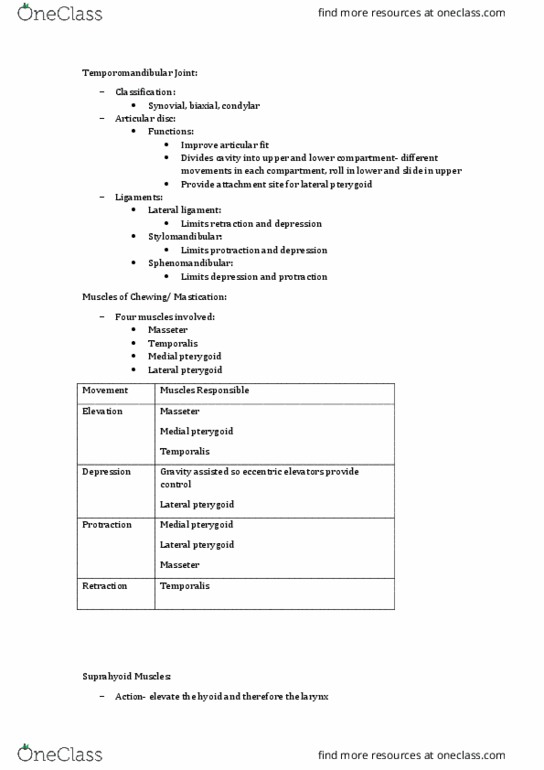
Articular surfaces: mandibular fossa of temporal bone, condyle of mandible. Articular disc: attaches to , condyle of mandible anteriorly, temporal bone posteriorly, articular capsule around its periphery, functions: Improve articular fit: divides cavity into upper and lower compartment- different movements in each compartment, roll in lower and slide in upper, provide attachment site for lateral pterygoid. Synovial cavity: two cavities, one below and one above disc. Chewing involves elevation and depression, and protraction and retraction of the mandible- in combinations. Four muscles involved: masseter, temporalis, medial pterygoid, lateral pterygoid. Not attached to any other part of the skeletal system, therefore very mobile. Positioned by musculature above (suprahyoid) and below (infrahyoid) Action- elevate the hyoid and therefore the larynx larynx and the sternum. Open or close the apertures of the face- mouth, nostrils, eyes. Circular muscles around the eye and mouth close these openings.