HUMB1000 Lecture Notes - Lecture 9: Arachnoid Mater, Medulla Oblongata, Dura Mater

12 Jun 2018
School
Department
Course
Professor

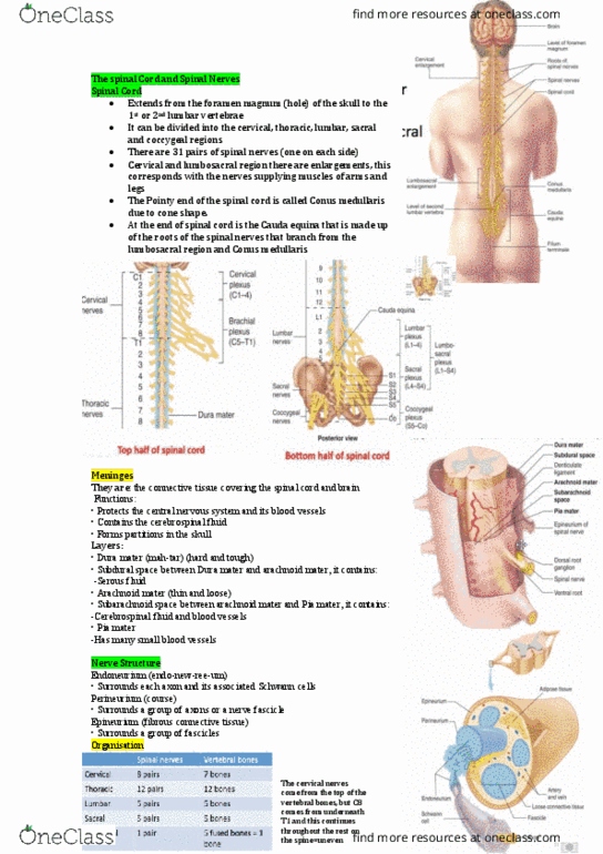
The Spinal Cord and Spinal Nerves
Spinal Cord
❖ Extends from the foramen magnum to the first or second lumbar vertebrae
❖ Can be divided into cervical, thoracic, lumbar, sacral and coccygeal regions
❖ 31 pairs of spinal nerves
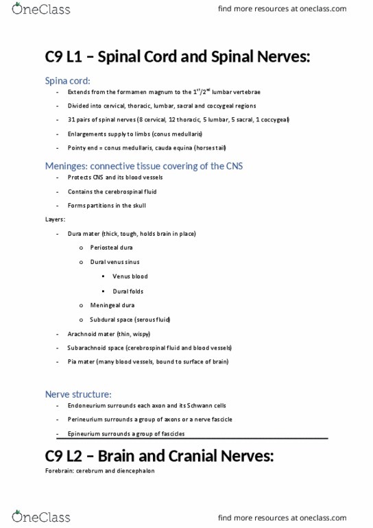
Meninges
❖ the connective tissue covering the spinal cord and brain
❖ Functions:
o Protects the central nervous system and its blood vessels
o Contains the cerebrospinal fluid
o Forms partitions in the skull
❖ Dura mater
o Subdural space
▪ Serous fluid
❖ Arachnoid mater
o Subararachnoid space
▪ Cerebrospinal and blood vessels
❖ Pia mater
o Has many small blood vessels
Organisation of neurons in the spinal cord and spinal nerves
❖ Sensory neurons travel through the dorsal roots
❖ Motor neurons (somatic and autonomic) neurons travel through the ventral roots
❖ Spinal nerves contain sensory neurons and motor (somatic and autonomic) neurons
❖ Cell bodies of motor neurons are in horns of grey matter
o Somatic motor neuron cell bodies in anterior (ventral) horn (motor horn)
o Autonomic motor neuron cell bodies in lateral horn
find more resources at oneclass.com
find more resources at oneclass.com

Nerve Structure
❖ Endoneurium
o Surrounds each axon and its associated Schwann cells
❖ Perineurium
o Surrounds a group of axons or a nerve fascicle
❖ Epineurium
o Surrounds a group of fascicles
Organisation of spinal nerves
Spinal Nerves
Vertebral Bones
Cervical
8
7
Thoracic
12
12
Lumbar
5
5
Sacral
5
5
Coccygeal
1
1
Where do sensory and motor nerve roots enter and leave the spinal cord?
find more resources at oneclass.com
find more resources at oneclass.com

The Brain and Cranial Nerves
The Brain
• Forebrain
o Cerebrum
o Diencephalon
• Midbrain
• Hindbrain
o Pons
o Medulla oblongata
o Cerebellum
Medulla Oblongata
❖ Autonomic reflex centre maintaining body homeostasis
❖ Cardiovascular centre
o Regulates heart rate, force of heart contraction and blood vessel diameter
❖ Respiratory centre
o Regulates rate and depth of breathing
❖ Other reflexes
o Swallowing, vomiting, hiccupping, coughing and sneezing
Pons
❖ Pons = bridge
❖ Contains conduction tracts:
o Longitudinal tracts from the spinal cord to higher brain centres
o Transverse tracts form the cerebrum (motor cortex) and cerebellum
❖ Sleep centre
o Rapid eye movement
❖ Respiratory centre
Midbrain
❖ Receives visual, auditory and tactile sensory input generating reflex movements of
the head, eyes and body
❖ Controlling movement of the eye
Cerebellum
❖ Cerebellum = little brain
❖ Controls locomotion, in association with the cerebrum
❖ Controls fine motor control
❖ Controls posture and balance
find more resources at oneclass.com
find more resources at oneclass.com
Document Summary
Extends from the foramen magnum to the first or second lumbar vertebrae. Can be divided into cervical, thoracic, lumbar, sacral and coccygeal regions. The connective tissue covering the spinal cord and brain. Functions: protects the central nervous system and its blood vessels, contains the cerebrospinal fluid, forms partitions in the skull. Dura mater: subdural space, serous fluid. Arachnoid mater: subararachnoid space, cerebrospinal and blood vessels. Pia mater: has many small blood vessels. Organisation of neurons in the spinal cord and spinal nerves. Sensory neurons travel through the dorsal roots. Motor neurons (somatic and autonomic) neurons travel through the ventral roots. Spinal nerves contain sensory neurons and motor (somatic and autonomic) neurons. Cell bodies of motor neurons are in horns of grey matter: somatic motor neuron cell bodies in anterior (ventral) horn (motor horn, autonomic motor neuron cell bodies in lateral horn. Endoneurium: surrounds each axon and its associated schwann cells. Perineurium: surrounds a group of axons or a nerve fascicle.