HUMB1000 Lecture Notes - Lecture 7: Distal Convoluted Tubule, Renal Capsule, Loose Connective Tissue

12 Jun 2018
School
Department
Course
Professor

Urine Formation
❖ Kidneys primary function is to filter blood and form urine
❖ Regulate fluid balance, electrolyte concentration, and pH excreting unwanted fluid
and substances as urine
❖ Glomerular filtration occurs as blood circulates through the glomerulus.
o Fluid, small molecules and ions in the blood within glomerular capillaries
move across the filtration membrane
❖ Once filtrate enters the renal tubule, it is called tubular fluid
❖ Substances are simultaneously removed from, and added to the tubular fluid as it
flows through the renal tubule
❖ In tubular reabsorption, useful substance is removed from the tubular fluid and
returned back into the blood
❖ Solutes move across the tubule wall into the interstitial fluid by processes such as
diffusion, facilitated diffusion, active transport, cotransport, and osmosis
❖ Water reabsorption
o Proximal convoluted tubule – 65%
o Loop of Henle (descending limb) – 15%
o Distal convoluted tubule/collecting ducts – 19%
❖ 99% of the glomerular filtrate ultimately returns to the bloodstream, 1% or less that
is not reabsorbed will be excreted as urine
➢ TUBULAR SECRETION – substances move from the blood capillary to the tubular fluid
o Water materials – metabolic by-products, excess ions and drugs – diffuse
out of the blood to the interstitial fluid, and are then transported across the
tubular wall and secreted into the tubular fluid for excretion
❖ ABILITY TO CONCENTRATE URINE:
o maintaining high concentration of solutes sodium chloride and urea in the
renal medulla
o presence of antidiuretic hormone (ADH) –makes collecting ducts more
permeable to water
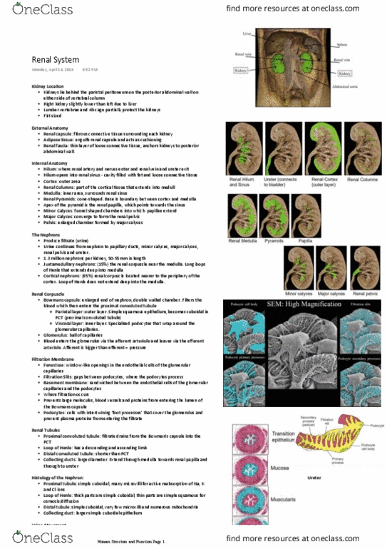
Sodium-Potassium Exchange Pump
❖ Moves sodium ions out of the cell and potassium ions into the cell
❖ 3 sodium ions and an ATP molecule can bind to the carrier protein on the inside of
the cell membrane
❖ ATP is broken down to ADP and phosphate, the carrier protein changes shape and
the sodium ions are transported across the membrane
❖ Potassium ions outside of the cell bind to the carrier protein
❖ 2 potassium ions move into the cell and the attached phosphate is released
❖ The protein now goes back to the original shape
find more resources at oneclass.com
find more resources at oneclass.com

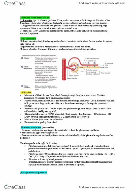
Cotransport (Symport and Antiport)
❖ Small molecules such as sugars and amino acids can be transported up a
concentration gradient
❖ The sugar moves via a membrane transport protein form
outside of the cell where the sugar concentration is low to
the inside of the cell where the sugar concentration is high
❖ The transport of the sugar through coupled transport
protein is driven by the movement of counter-ions such as
sodium or protons moving down their concentration
gradient, from a region of high to low concentration
❖ Sodium ions and the specific sugar or amino acid
simultaneously bind to the same transmembrane protein
on the outside of the cell, called a SYMPORT
❖ When the counter-ion is sodium, the low concentration of
sodium on the inside of the cell required to transport the
sugar is maintained by the sodium/potassium pump
❖ COUNTER-TRANSPORT – inward movement of sodium ions
is coupled with the outer movement of another substance
such as calcium ions
❖ As in co-transport, the sodium ions and the other
substance bind to the same transport protein, called the
ANTIPORT. They bind on opposite sides of the membrane
and are moved in opposite directions
find more resources at oneclass.com
find more resources at oneclass.com

Anatomy of the Renal System
Kidney Location
❖
❖ The kidneys lie behind the parietal peritoneum on the posterior abdominal wall on
either side of vertebral column.
❖ Right kidney slightly lower than left (due to liver)
❖ Lumbar vertebrae and rib cage partially protect the kidneys
❖ Bean shaped
❖ Size of a fist (~130g)
Kidney Location & External Anatomy
❖ Renal capsule: fibrous connective tissue surrounding each kidney.
❖ Adipose tissue: engulfs renal capsule and acts as cushioning
❖ Renal fascia: thin layer loose connective tissue, which anchors kidneys to posterior
abdominal wall.
Kidney External Anatomy
❖ Hilum: Renal artery and nerves enter and renal vein
and ureter exit
❖ Hilum opens into renal sinus: cavity filled with fat
and loose connective tissue
❖ Ureter: exits at the hilum; connects to urinary
bladder
❖ Cortex: outer area
❖ Renal columns: part of cortical tissue that extends
into medulla
Kidney Internal Anatomy
❖ Medulla: inner area, surrounds renal sinus
❖ Renal pyramids: cone-shaped. Base is
boundary between cortex and medulla.
❖ Apex of pyramid is renal papilla, points
toward sinus.
❖ Minor Calyces: funnel shaped chambers into
which papillae extend.
❖ Major Calyces: converge to form the renal
pelvis
❖ Pelvis: enlarged chamber formed by major
calyces
find more resources at oneclass.com
find more resources at oneclass.com
Document Summary
Kidneys primary function is to filter blood and form urine. Regulate fluid balance, electrolyte concentration, and ph excreting unwanted fluid and substances as urine. Glomerular filtration occurs as blood circulates through the glomerulus: fluid, small molecules and ions in the blood within glomerular capillaries move across the filtration membrane. Once filtrate enters the renal tubule, it is called tubular fluid. Substances are simultaneously removed from, and added to the tubular fluid as it flows through the renal tubule. In tubular reabsorption, useful substance is removed from the tubular fluid and returned back into the blood. Solutes move across the tubule wall into the interstitial fluid by processes such as diffusion, facilitated diffusion, active transport, cotransport, and osmosis. Water reabsorption: proximal convoluted tubule 65, loop of henle (descending limb) 15, distal convoluted tubule/collecting ducts 19% 99% of the glomerular filtrate ultimately returns to the bloodstream, 1% or less that is not reabsorbed will be excreted as urine.