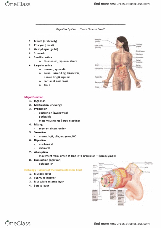
HUMB1001 Lecture Notes - Lecture 5: Loose Connective Tissue, Lamina Propria, Basement Membrane


How is blood circulation regulated?
Systemic Circulation
- Systemic circulation (84%)
o blood to the body
- Heart chambers (7%)
o blood to the heart muscles
- Pulmonary vessels (9%)
o blood to and from lungs
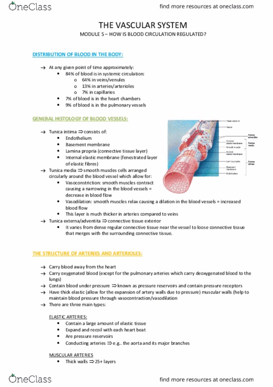
Histology of arteries & veins
Tunica intima
• Endothelium – simple squamous layer
• Basement membrane
• Lamina propria (Connective Tissue [C.T.]
layer)
• Internal elastic membrane. Fenestrated layer
of elastic fibres.
Tunica media
• Smooth muscle cells arranged circularly
around the blood vessel
• Vasoconstriction: smooth muscles, contract,
decrease in blood flow
• Vasodilation: smooth muscles relax, increase in blood flow
Tunica externa (adventitia)
• Connective tissue
• Varies from dense regular near the vessel to loose; that merges with the
surrounding C.T.
Arteries & arterioles
Elastic arteries
e.g. aorta & major branches
• Elastic tissue
• expand and recoil
• are pressure reservoirs
• Conducting arteries
Muscular arteries
e.g. medium size arteries
• Thick walls – 25+ layers of muscle
• Undergo vasoconstriction & vasodilation
• Distributing arteries
Arterioles
e.g. small muscular arteries
• Decreasing in size with gradual loss of wall layers down to terminal arterioles
veins/venules 64%
arteries/arterioles 13%
capillaries 7%
find more resources at oneclass.com
find more resources at oneclass.com

Venules & veins
Venules
• Very small veins that drain capillary network
• Endothelial cells and basement membrane with a
few smooth muscle cells
• As diameter of venules increases, amount of
smooth muscle increases
Veins
• Smooth muscle cells form continuous layer; addition of tunica
adventia
• Have valves (to prevent back flow), thin walls & large lumens
• Are very compliant (obey rules)
o 24 x more compliant than arteries
• Capacitance vessels of circulation
o i.e. hold a lot of blood at very low pressure
Artery Vs Vein
find more resources at oneclass.com
find more resources at oneclass.com

Capillaries
• Very small vessels (<10 microns)
• Endothelial cells sitting on a basement membrane & delicate layer of loose
connective tissue
• Designed for rapid exchange of nutrients & metabolites between blood & interstitial
fluid
• Substances move through capillary wall via diffusion:
o Lipid-soluble and small water-soluble molecules through plasma membrane
o Large water-soluble molecules pass through fenestrae or gaps between
endothelial cells
Three types of capillaries:
Continuous
• No gaps between endothelial cells
o e.g. muscle, sin
Fenestrated
• Highly permeable
o e.g. kidney, endocrine glands
Sinusoids
• Large diameter & large fenestrae
o e.g. liver, bone marrow, spleen
find more resources at oneclass.com
find more resources at oneclass.com
Document Summary
Systemic circulation (84%: blood to the body. Heart chambers (7%: blood to the heart muscles. Pulmonary vessels (9%: blood to and from lungs. Tunica intima: endothelium simple squamous layer, basement membrane. Tunica media: smooth muscle cells arranged circularly around the blood vessel, vasoconstriction: smooth muscles, contract, decrease in blood flow, vasodilation: smooth muscles relax, increase in blood flow. Tunica externa (adventitia: connective tissue, varies from dense regular near the vessel to loose; that merges with the surrounding c. t. Elastic arteries e. g. aorta & major branches: elastic tissue, expand and recoil, are pressure reservoirs, conducting arteries. Muscular arteries e. g. medium size arteries: thick walls 25+ layers of muscle, undergo vasoconstriction & vasodilation, distributing arteries. Arterioles e. g. small muscular arteries: decreasing in size with gradual loss of wall layers down to terminal arterioles. Venules: very small veins that drain capillary network, endothelial cells and basement membrane with a few smooth muscle cells, as diameter of venules increases, amount of smooth muscle increases.