MMED1005 Lecture Notes - Lecture 19: Pulmonary Function Testing, Asthma, Elastin

7 Jun 2018
School
Department
Course
Professor

Respiratory lecture 2: mechanics of breathing
- The thoracic cavity
o The lungs
o The pleural cavity
- Elastic recoil of the lung
- The muscles involved in breathing
o Main: Diaphragm
o Also, the accessory muscles
- Pressure changes in the lung
- Spirometry
o Lung volumes
Revision of the previous lecture:
- Conducting portion:
o Nasal cavities
o Pharynx
o Larynx
o Trachea
o Bronchi
o Terminal bronchioles
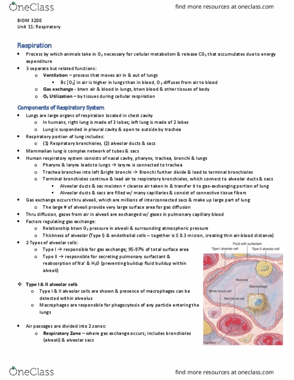
- Respiratory portion: only function is gas exchange
o Respiratory bronchioles
o Alveolar ducts
o Respiratory sac alveoli
- Thin membrane between alveoli wall and capillaries – respiratory membrane
o This is where the gases are exchanged
o Oxygen higher pressure in alveolus → venous blood, becoming arteriole
o Carbon dioxide goes in the opposite direction
o
find more resources at oneclass.com
find more resources at oneclass.com

The thoracic cavity:
- around each lung, there is the pleural cavity. They separately seal each lung
o if one lung fails, the other wont be affected
- two membranes:
o visceral pleura: stuck to lung
o parietal pleura: stuck to ribs
- intrapleural fluid: keeps the membranes together
Pressure in the lungs and pleural cavities:
- the intrapleural pressure is lower than 760mmg negative pressure of -4mmHg
- the lung want to collapse and the ribs want to bow outward
- two opposing forces which create the negative pressure in pleural cavity: elastic
recoil of the lung and elastic recoil of the rib cage
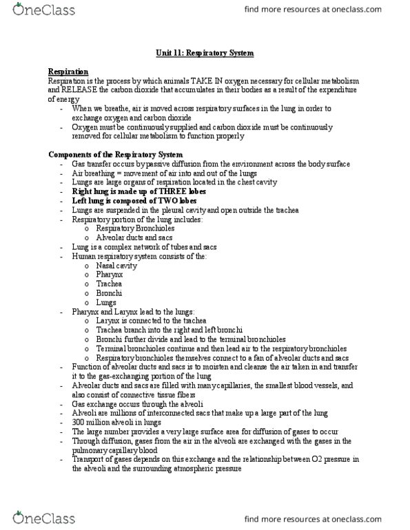
The thoracic activity:
- first force: elastic recoil of the lungs
o natural tendency of the lung is to collapse
because of its elastic recoil
- second force: elastic recoil of the chest wall (rib
cage)
o tends to expand
- these two forces pull apart sealed pleural cavity,
creating negative pressure
- at rest, these two forces are in equilibrium and
negative intrapleural pressure keeps lung partially
expanded between breath
find more resources at oneclass.com
find more resources at oneclass.com
Document Summary
The thoracic cavity: the lungs, the pleural cavity. The muscles involved in breathing: main: diaphragm, also, the accessory muscles. Conducting portion: nasal cavities, pharynx, larynx, trachea, bronchi, terminal bronchioles. Respiratory portion: only function is gas exchange: respiratory bronchioles, alveolar ducts, respiratory sac alveoli. Thin membrane between alveoli wall and capillaries respiratory membrane: this is where the gases are exchanged, oxygen higher pressure in alveolus venous blood, becoming arteriole, carbon dioxide goes in the opposite direction. Around each lung, there is the pleural cavity. They separately seal each lung if one lung fails, the other wont be affected two membranes: visceral pleura: stuck to lung, parietal pleura: stuck to ribs intrapleural fluid: keeps the membranes together. At rest, these two forces are in equilibrium and negative intrapleural pressure keeps lung partially expanded between breath. Muscles of respiration: in the resting state, diaphragm works, with some participation of external intercostal muscles.