BM 1041:03 Lecture Notes - Lecture 23: Olecranon Fossa, Trochlear Notch, Coronoid Fossa Of The Humerus

23 May 2018
School
Department
Course
Professor

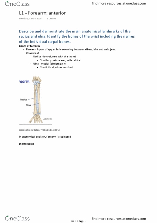
Describe and demonstrate the main anatomical landmarks of the
distal humerus, proximal radius and proximal ulna.
Distal
humerus
Screen clipping taken: 1/05/2018 6:45 PM
Bone flattens in anteroposterior plane
-
Lateral supracondylar ridge
-
Medial supracondylar ridge
-
Capitulum - articulates with radius of forearm
○
Trochlea - articulates with ulna of forearm, extends onto posterior
surface of humerus
○
2 articular parts
•
Condyle (blue part)
-
Oval impression for attachment of muscles in anterior compartment of
forearm
○
Ulnar nerve passes around its posterior surface of medial epicondyle,
can be palpated here
○
Medial epicondyle - major palpable landmark on medial side of elbow
•
Larger impression for attachment of posterior compartment muscles of
the forearm
○
Lateral epicondyle - less pronounced
•
2 epicondyles
-
Coronoid fossa
•
Radial fossa
•
Olecranon fossa
•
3 fossae
-
Proximal
L2 - Brachium and elbow
Monday, 30 April 2018
7:08 PM
wk 10 Page 1

Proximal
radius
Screen clipping taken: 1/05/2018 6:58 PM
Thick disc shaped
•
Horizontal plane
•
Concave for articulation with the capitulum of humerus
•
Thick margin of disc - broad medially to articulate with radial notch on prox
end of ulna
•
Head
-
Short and narrow
•
Between head and radial tuberosity on shaft
•
Neck
-
Large blunt projection on medial surface
•
Roughened surface for attachment of biceps brachii tendon
•
Radial tuberosity
-
Proximal
ulna
Screen clipping taken: 1/05/2018 6:52 PM
Projection, extends proximally
•
Superior surface marked by large impression for attachment of the triceps
brachii muscle
•
Olecranon
-
Projects anteriorly from proximal end
•
Articulates with olecranon in forming trochlear notch
•
Lateral surface marked by radial notch for articulation of head of radius
•
Coronoid process
-
Supinator crest
-
Radial notch
-
Articulates with trochlea of humerus
•
Trochlear notch
-
largest of the roughenings for muscle attachment
•
At the apex of the anterior surface
•
Attachment site for brachialis muscle
•
Ulnar tuberosity
-
wk 10 Page 2

Describe the anatomy of the elbow joint and the ligaments and their
attachments and function.
Elbow joint
Synovial hinge joint
-
Screen clipping taken: 1/05/2018 7:44 PM
Humerus
○
Ulna
○
Radius
○
Bones
-
Between trochlear notch of ulna and trochlea of humerus, and between head of
radius and the capitulum of humerus (principal articulations of elbow joint,
flex/ext)
▪
Uniaxial joint
▪
Humeroulnar
○
Joint between head of radius and radial notch of ulna, proximal radio-ulnar joint ->
supination and pronation of forearm
▪
Proximal radioulnar joint
○
Articular surfaces of bones covered with hyaline cartilage
○
3 separate articulations, which share a common synovial cavity
-
Screen clipping taken: 1/05/2018 7:44 PM
Articular capsule loose -> allow good range of flexion and extension
-
Separated from fibrous membrane by pads of fat in regions overlying the fossae
Synovial membrane originates from edges of articular cartilage and lines the radial fossa,
coronoid fossa, olecranon fossa, deep surface of the joint capsule, medial surface of the
trochlea
-
wk 10 Page 3
Document Summary
Describe and demonstrate the main anatomical landmarks of the distal humerus, proximal radius and proximal ulna. Trochlea - articulates with ulna of forearm, extends onto posterior surface of humerus. Medial epicondyle - major palpable landmark on medial side of elbow. Oval impression for attachment of muscles in anterior compartment of forearm. Ulnar nerve passes around its posterior surface of medial epicondyle, can be palpated here. Larger impression for attachment of posterior compartment muscles of the forearm. Concave for articulation with the capitulum of humerus. Thick margin of disc - broad medially to articulate with radial notch on prox end of ulna. Roughened surface for attachment of biceps brachii tendon. Superior surface marked by large impression for attachment of the triceps brachii muscle. Lateral surface marked by radial notch for articulation of head of radius. Ulnar tuberosity largest of the roughenings for muscle attachment. Attachment site for brachialis muscle wk 10 page 2.