BM 1041:03 Lecture Notes - Lecture 24: Distal Radioulnar Articulation, Extensor Pollicis Longus Muscle, Ulnar Styloid Process

23 May 2018
School
Department
Course
Professor

Describe and demonstrate the main anatomical landmarks of the
radius and ulna. Identify the bones of the wrist including the names
of the individual carpal bones.
Forearm is part of upper limb extending between elbow joint and wrist joint
-
Smaller proximal end, wider distal
▪
Radius - lateral, runs with the thumb
○
Small distal, wider proximal
▪
Ulna - medial (ulnderneath)
○
Consists of
-
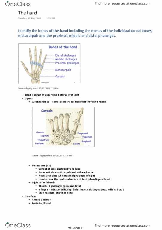
Bones of forearm
Screen clipping taken: 7/05/2018 1:23 PM
In anatomical position, forearm is supinated
Distal radius
L1 - Forearm: anterior
Monday, 7 May 2018
1:20 PM
wk 11 Page 1

Screen clipping taken: 7/05/2018 1:28 PM
Anterior border
○
Posterior border
○
Interosseous border
○
3 borders
-
Anterior, posterior, lateral
○
3 surfaces
-
Oblique line, near roughened area for pronator teres
○
Radial styloid process - insertion of brachioradialis and radial collateral ligament)
○
Ulnar notch - articulation with ulna
○
Anterior border - sharp ridge forming lateral margin
○
Large anterior and posterior surfaces, narrow medial and lateral surfaces
○
Anterior view
-
Dorsal tubercle - helps keep extensor pollicis longus in place, acts as pulley for tendon of
extensor muscle of thumb
○
Posterior view
-
Facet of scaphoid - articulates with scaphoid carpal bone
○
Facet for lunate - articulates with lunate
○
Both contribute to radiocarpal (wrist) joint
○
Inferior view
-
Distal ulna
wk 11 Page 2

Screen clipping taken: 7/05/2018 1:34 PM
Anterior, posterior, lateral
○
3 surfaces
-
Anterior, posterior, interosseous
○
3 borders
-
Ulnar styloid process - attached to ulnar collateral ligament and palmar ulnocarpal ligament
-
Facet for articular disc - for fibrocartilaginous disc
-
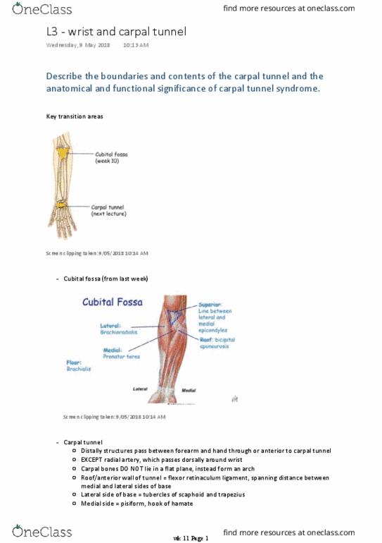
Some lovers try positions that they cant handle
○
Prox row: scaphoid lunate triquetral pisiform
○
Dist row: trapezius trapezoid capitate hamate
○
Screen clipping taken: 9/05/2018 7:52 PM
8 carpal bones = bones of wrist
-
6 metacarpals = bones of metacarpus
-
Phalanges = bones of the digits (thumb has 2, rest of digits has 3)
-
Bones of wrist
Describe the anatomy of the proximal and distal radio‐ulnar joints.
wk 11 Page 3
Document Summary
Describe and demonstrate the main anatomical landmarks of the radius and ulna. Identify the bones of the wrist including the names of the individual carpal bones. Forearm is part of upper limb extending between elbow joint and wrist joint. Oblique line, near roughened area for pronator teres. Radial styloid process - insertion of brachioradialis and radial collateral ligament) Anterior border - sharp ridge forming lateral margin. Large anterior and posterior surfaces, narrow medial and lateral surfaces. Dorsal tubercle - helps keep extensor pollicis longus in place, acts as pulley for tendon of extensor muscle of thumb. Facet of scaphoid - articulates with scaphoid carpal bone. Ulnar styloid process - attached to ulnar collateral ligament and palmar ulnocarpal ligament. Facet for articular disc - for fibrocartilaginous disc. Some lovers try positions that they cant handle. Phalanges = bones of the digits (thumb has 2, rest of digits has 3)