BM 1041:03 Lecture Notes - Lecture 20: Glenoid Labrum, Coracoacromial Ligament, Biceps

23 May 2018
School
Department
Course
Professor

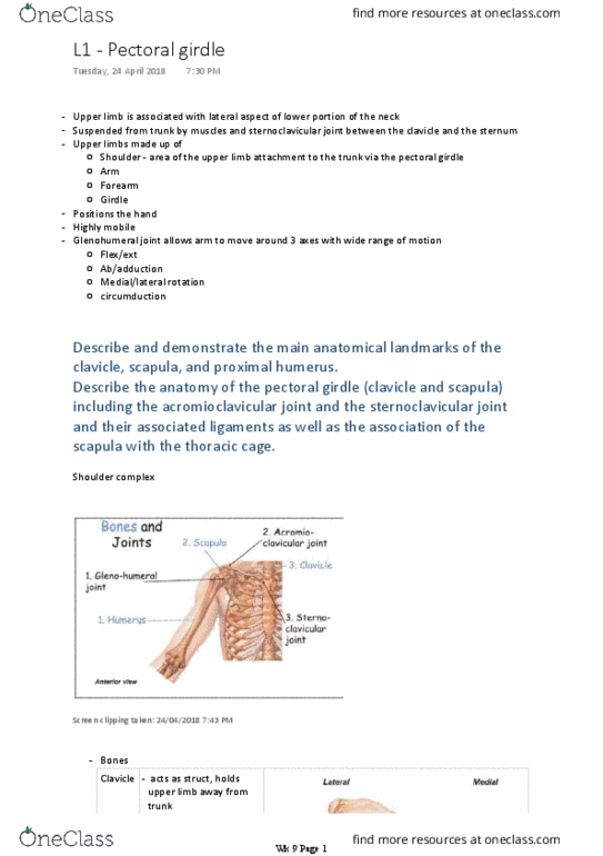
Describe the anatomy of the gleno‐humeral joint including the
factors that contribute to the movement and stability of the
glenohumeral joint: the rotator cuff muscles, the long head of biceps
brachii and the specific ligaments. Based on this anatomy explain
the functional and clinical consequences of a dislocation of the
glenohumeral dislocation
GH joint
Ball and socket between head of humerus and glenoid cavity of scapula
-
Flex/ext, ab/adduction, med/lat rotation, circumduction
○
Multiaxial - large range of movement
-
L2 - The shoulder
Wednesday, 25 April 2018
11:28 AM
Wk 9 Page 1

Screen clipping taken: 25/04/2018 3:28 PM
Poor bony congruity
-
Stability provided by rotator cuff muscles, long head of biceps brachii, related bony
processes and extracapsular ligaments
○
Stability provided by surrounding muscle tendons and skeletal arch formed superiorly by
coracoid process and acromion and coracoacromial ligament
○
Holds head of humerus in glenoid cavity
▪
Tendons of rotator cuff muscles blend with joint capsule to forma musculotendinous
collar that surrounds posterior, superior and anterior aspects of joint
○
Tendon of long head of biceps brachii passes superiorly through joint - restricts upward
movement of humeral head on glenoid cavity
○
Mobility > stability (sacrificed)
-
Large spherical head of the humerus
○
Deepened and expanded peripherally by fibrocartilaginous collar (glenoid labrum),
which attaches to margin of fossa
▪
Small glenoid cavity of scapula
○
Surfaces covered by hyaline cartilage
○
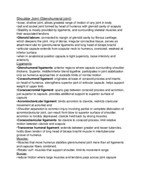
Articular surfaces
-
Glenoid labrum
○
Synovial membrane attaches to margins of articular surfaces and lines fibrous
membrane of joint capsule
▪
Loose and thin inferiorly, accommodates for abduction of arm
▪
Inferior part is weakest - only part NOT reinforced by rotator cuff -> prone to
dislocation
▪
Articular capsule
○
Weak
▪
Extrinsic and superior - coracohumeral ligament
▪
Intrinsic and anterior - glenohumeral ligament
▪
Very strong, prevents superior displacesment
□
Coracoacromial
▪
Ligaments
○
Features
-
Wk 9 Page 2

Rotator cuffs
▪
Long head of biceps brachii
▪
Muscle tendons (major stabilisers)
○
Sacs of synovial fluid
▪
Synovial membrane protrudes through apertures in fibrous membrane to form
bursae
▪
Reduce friction between tendons and adjacent joint capsule and bone
▪
Lies between subscapularis and fibrous membrane
□
Subtendinous bursa of subscapularis
▪
Between acromion and supraspinatus
□
Subacromial bursa
▪
Bursae
○
Screen clipping taken: 25/04/2018 3:14 PM
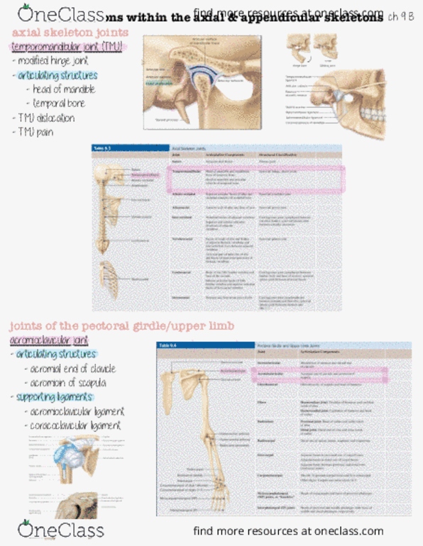
attachments
Function
Rotator
cuff
muscles
(4)
Supraspinatus
•
Infraspinatus
•
Teres minor
•
3 posterior rotator cuff attach to greater tubercle
-
Subscapularis attaches to lesser tubercle
-
Keep humeral head
centred within glenoid
cavity of scapula, while
powerful muscles lift
arm
-
Muscles nmust be in
-
Wk 9 Page 3
Document Summary
Describe the anatomy of the gleno hu(cid:373)eral joi(cid:374)t i(cid:374)cludi(cid:374)g the factors that contribute to the movement and stability of the glenohumeral joint: the rotator cuff muscles, the long head of biceps brachii and the specific ligaments. Based on this anatomy explain the functional and clinical consequences of a dislocation of the glenohumeral dislocation. Ball and socket between head of humerus and glenoid cavity of scapula. Stability provided by rotator cuff muscles, long head of biceps brachii, related bony processes and extracapsular ligaments. Stability provided by surrounding muscle tendons and skeletal arch formed superiorly by coracoid process and acromion and coracoacromial ligament. Tendons of rotator cuff muscles blend with joint capsule to forma musculotendinous collar that surrounds posterior, superior and anterior aspects of joint. Tendon of long head of biceps brachii passes superiorly through joint - restricts upward movement of humeral head on glenoid cavity. Deepened and expanded peripherally by fibrocartilaginous collar (glenoid labrum), which attaches to margin of fossa.