ANAT20006 Lecture Notes - Lecture 3: Paraxial Mesoderm, Intermediate Mesoderm, Yolk Sac

12 Jun 2018
School
Department
Course
Professor

Lecture 2 - Wednesday 26 July 2017
ANAT20006 - HUMAN STRUCTURE & FUNCTION
LECTURE 3
EMBRYOLOGY (2)
LECTURE 2:
•
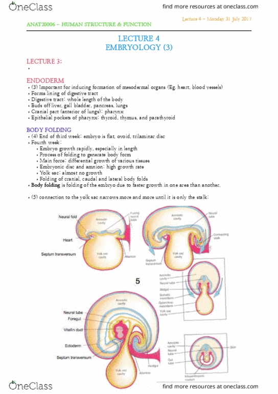
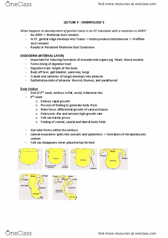
BODY FOLDING
•End of third week: embryo is flat, ovoid, trilaminar disc. It begins growing
really fast. Ectoderm grows faster than endoderm.
•Fourth week:
•Embryo growth rapidly, especially in length
•Process of folding to generate body form
•Main force: differential growth of various tissues
•Embryonic disc and amnion: high growth rate
•Yolk sac: almost no growth
•Developing notochord, neural tube and somites stiffen dorsal axis
•Folding of cranial, caudal and lateral body folds
•

Lecture 2 - Wednesday 26 July 2017
ANAT20006 - HUMAN STRUCTURE & FUNCTION
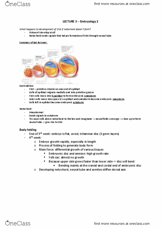
EMBRYO
FOLDING
•Intra
embryonic
cavity =
coelom.
•Transverse sections.
•Disc with ecto endo and
mesoderm.
•The red layer is the mesoderm. In
B it swells up a bit and we can
see that the neural tube forms.
•

Lecture 2 - Wednesday 26 July 2017
ANAT20006 - HUMAN STRUCTURE & FUNCTION
MESODERM
•Divided into:
•Paraxial mesoderm
•Intermediate mesoderm
•Lateral mesoderm
•Each gives rise to different organs within the embryo.
PARAXIAL MESODERM
•Dermis of skin
•Axial skeleton
•Axial and limb muscles
INTERMEDIATE MESODERM
•Urogenital system
LATERAL MESODERM
•Divided into somatic and splanchnic
•Ventro-lateral body wall (connective
tissue, not muscle)
•Heart and vasculature
•Wall of gut
•Bones of the limbs
PARAXIAL MESODERM
•In the head region:
•Head mesoderm (together with neural
crest) forms skeleton, muscles and
connective tissue of face and skull
•In trunk region:
•Forms somites, which will produce
muscle, bone, and dermis. This uses
EMT.
•(do not mix up mesenchymal and
mesodermal cells)
SOMITOGENESIS (1)
•Epithelialization
•Fissure formation (separation)
•Periodicity
•Specification
•Differentiation
•End up with a ball of some
epithelial and some mesenchymal !
Document Summary
Body folding: end of third week: embryo is flat, ovoid, trilaminar disc. Folding: intra embryonic cavity = coelom, transverse sections, disc with ecto endo and mesoderm, the red layer is the mesoderm. B it swells up a bit and we can see that the neural tube forms. Mesoderm: divided into, paraxial mesoderm, intermediate mesoderm, lateral mesoderm, each gives rise to different organs within the embryo. Paraxial mesoderm: dermis of skin, axial skeleton, axial and limb muscles. Lateral mesoderm: divided into somatic and splanchnic, ventro-lateral body wall (connective tissue, not muscle, heart and vasculature, wall of gut, bones of the limbs. Paraxial mesoderm: in the head region, head mesoderm (together with neural crest) forms skeleton, muscles and connective tissue of face and skull, in trunk region, forms somites, which will produce muscle, bone, and dermis. Emt: (do not mix up mesenchymal and mesodermal cells)