ANAT20006 Lecture Notes - Lecture 29: Renal Fascia, Adrenal Gland, Abdominal Wall

1 Feb 2020
School
Department
Course
Professor
Document Summary
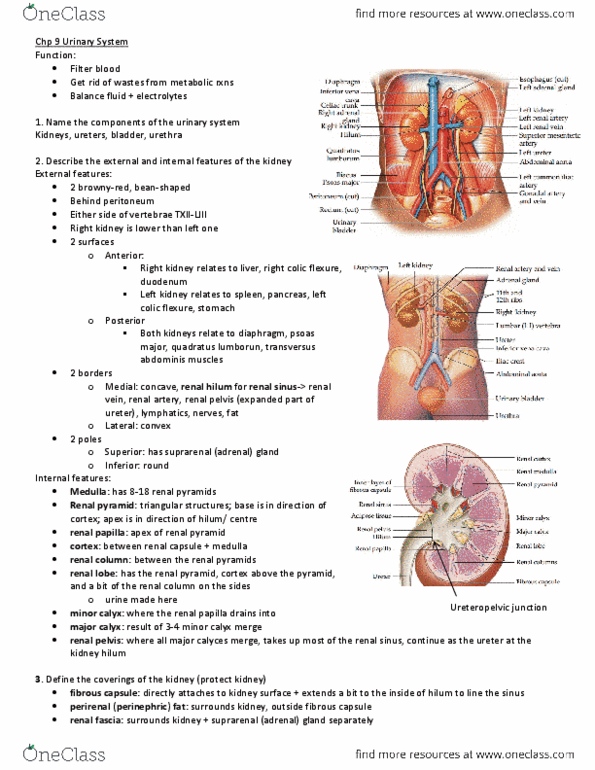
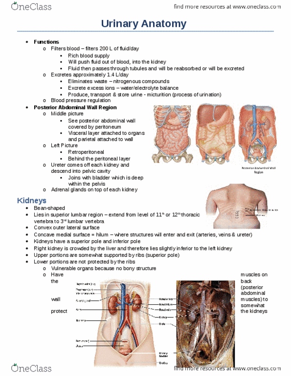
Lecture 29 urinary system and kidney (11. 1) Adrenal gland sits on top of kidney: part of endocrine system not part of urinary system. Retroperitoneal: anterior surface partially covered by peritoneum, no mesenteries. 2 surfaces: anterior surface, posterior surface sitting against posterior abdominal wall. When operating: can approach it laterally or posteriorly, can remove part of rib. 11/12: don"t need to interfere with peritoneal cavity. Kidney hilum tilts slightly forward anteromedial. Perinephric fat surrounds surface of kidney + goes into hilum. Renal sinuses are filled by perinephric fat. Vein is most anterior -> renal artery -> renal pelvis is most posterior. Renal artery comes off from abdominal aorta as paired structure. Right kidney is slightly lower than left (liver sits right above) Ivc receives blood from left and right renal vein: right is direct drainage, left has to pass in front of abdominal aorta -> drain into ivc, slightly longer because its further away has to pass midline.