ANAT30007 Lecture 28: [H1/91 notes] Vessels of Lower Limb A

10 Jun 2018
School
Department
Course
Professor

10.1 Vessels of Lower Limb
Monday, 11 May 2015
11:58 PM
Locomotor Page 1

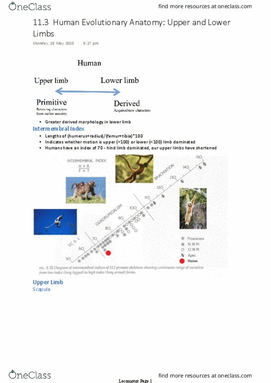
Abdominal aorta > common iliac > internal/external iliac
•
ant/post femoral circumflex (ant thigh muscles)
▪
Perforating vessels through adductor magnus to supply hamstrings
▪
Profunda femoris on adductor magnus
○
External iliac goes under inguinal ligament > femoral artery
•
Genicular arteries: anastomose around knee joint
○
Femoral artery through adductor hiatus > popliteal artery
•
Anterior tibial > under extensor retinaculum > dorsalis pedis > pierces first web space >
sole of foot
○
Gives off fibular artery to lateral compartment
▪
Goes behind medial malleolus to sole of foot > medial and lateral plantar
▪
Lateral plantar swings laterally then turns to create arch - connects with dorsalis
pedis
▪
Posterior tibial
○
At inferior border of popliteus, bifurcates into
•
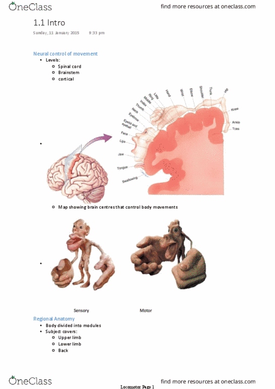
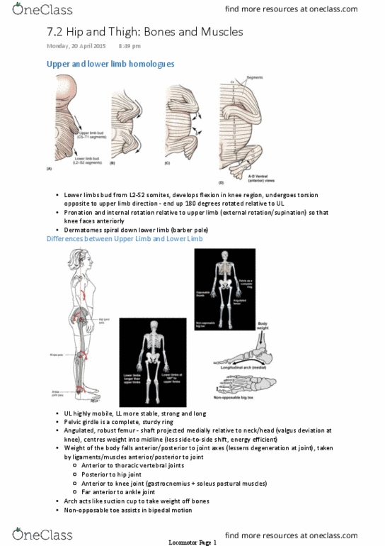
Contains artery, vein, femoral canal (contains deep inguinal lymph nodes/vessels and
fat)
○
Femoral canal has a funnel shape: femoral ring forms mouth, then blends with
adventitia of vessels
○
Boundaries of femoral ring: lateral vein, medial lacunar ligament, anterior inguinal
ligament, posterior floor of femoral triangle = pectineus
○
Femoral sheath
•
Locomotor Page 2
Document Summary
Abdominal aorta > common iliac > internal/external iliac. External iliac goes under inguinal ligament > femoral artery. Profunda femoris on adductor magnus ant/post femoral circumflex (ant thigh muscles) Perforating vessels through adductor magnus to supply hamstrings. Femoral artery through adductor hiatus > popliteal artery. Anterior tibial > under extensor retinaculum > dorsalis pedis > pierces first web space > sole of foot. Goes behind medial malleolus to sole of foot > medial and lateral plantar. Lateral plantar swings laterally then turns to create arch - connects with dorsalis pedis. Contains artery, vein, femoral canal (contains deep inguinal lymph nodes/vessels and fat) Femoral canal has a funnel shape: femoral ring forms mouth, then blends with adventitia of vessels. Boundaries of femoral ring: lateral vein, medial lacunar ligament, anterior inguinal ligament, posterior floor of femoral triangle = pectineus. Locomotor page 2 ligament, posterior floor of femoral triangle = pectineus.