ANAT30008 Lecture Notes - Lecture 9: Thoracolumbar Fascia, Renal Fascia, Psoas Minor Muscle

16 Oct 2018
School
Department
Course
Professor
Document Summary
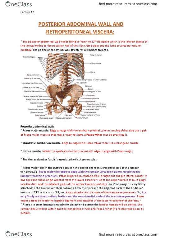
Lecture 9 posterior abdominal wall and retroperitoneal viscera. Bony framework of posterior abdominal wall = 12th ribs,t12, l1-5 and iv discs and pelvis. Perinephric fat and renal fascia: hilum of kidney is anteromedial, renal fascia encloses kidneys, hilum and great vessels in region, perinephric fat is between kidney and renal fascia, and goes into the hilum/renal sinuses. Renal pelvis (pass through renal hilum) ureter. Note: each kidney has 5 segments that have independent blood supply and thus is surgically resectable. Note: kidney initially developed in pelvis, then moved up hence why accessory renal artery stems from the aorta lower down (only present in 25% of people, not necessary once kidneys move up had renal arteries are developed) Ureter: muscular tube ~25cm long, renal pelvis (wider, behind renal artery) descend in front of psoas major and pelvic ring aligned with transverse processes of vertebrae .