ANAT30008 Lecture Notes - Lecture 29: Supraorbital Foramen, Zygomatic Bone, Macular Degeneration

16 Oct 2018
School
Department
Course
Professor
Document Summary
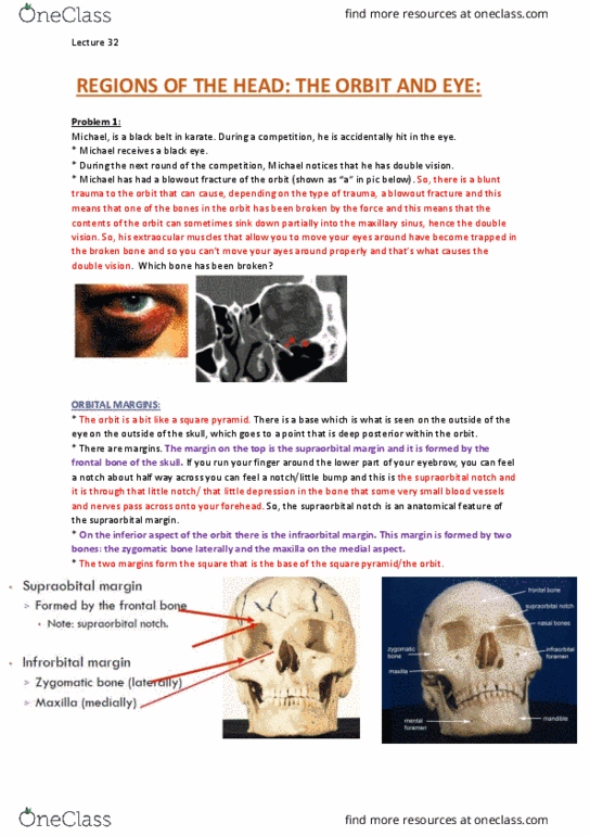
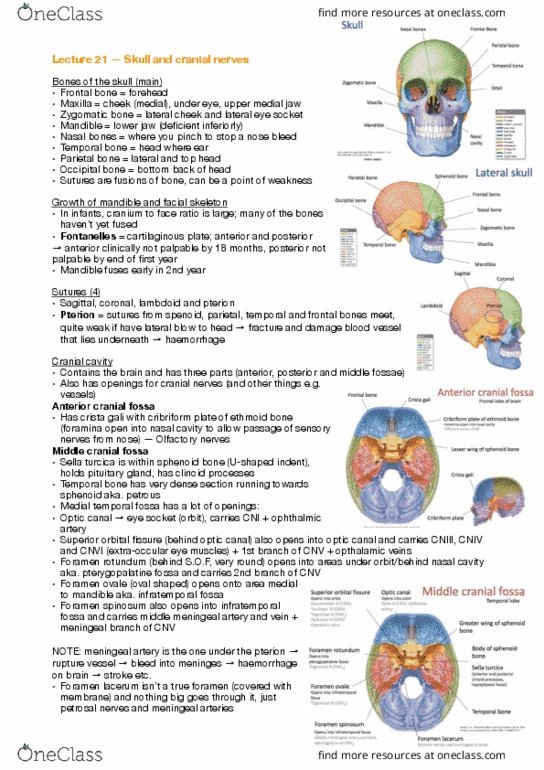
During a competition, he is accidentally hit in the eye. During the next round of the competition, michael notices that he has double vision. Had blunt trauma to the eye, one of the weakest orbital bones was broke = blowout fracture of the orbit. Supraorbital margin frontal bone note: supraorbital notch. Infraorbital margins zygomatic bone (laterally) & maxilla (medially) Roof = frontal bone, lesser wing of sphenoid. Floor = maxilla, zygomatic and palatine (tiny pink one) Lateral wall = zygomatic, greater wing of sphenoid. Medial wall = maxilla, lacrimal bone, ethmoid, body of sphenoid. Note: lacrimal bone very fragile and ethmoid bone has an area of paper-thin weakness most common bone broken in blowout fractures. Optic canal in sphenoid optic nerve (cnii) Superior orbital ssure in sphenoid cniii, cniv, cnvi, cnv. Orbital contents eyeball: outer coat cornea and sclera, function = strength. Cornea = front clear layer when contact lenses sit.