BIOL10004 Lecture Notes - Lecture 14: Extracellular Fluid, Cephalopod, Annelid

10 Jun 2018
School
Department
Course
Professor

Why do animals have circulatory systems?
Transfer of O2 and nutrients
-
Waste products removed
-
Communication via hormones
-
Temp regulation + reproduction
-
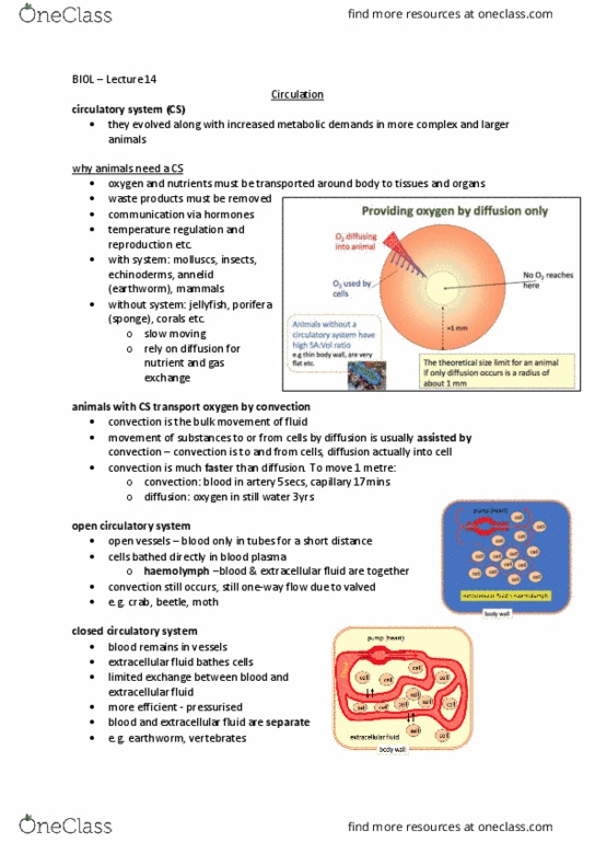
Animals w/o rely on diffusion - small and thin
Jellyfish
○
Sponge
○
Flatworm
○
Corals and sea anemones
○
HIGH SA:VOLUME RATIO for diffusion
○
-
Animals w system transport by convection
Convection: bulk flow of a fluid (air or water)
Movement assisted by convection - faster than diffusion
§
○
-
Open vs. closed systems
Both contain pumps - heart
-
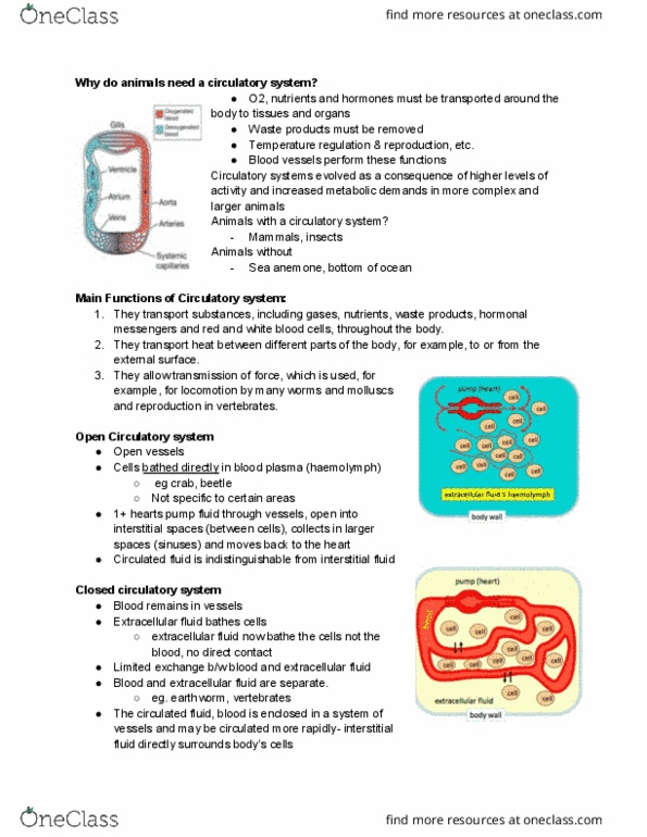
Open
In many invertebrates (eg. Crustaceans and some molluscs)
○
Heart pumps fluid through vessels that open into INTERSTITIAL SPACES
○
Fluid spreads among cells -> slowly returns to heart - often by gills that
oxygenate fluid on return
○
Cant distinguish circulated fluid from interstitial fluid
○
-
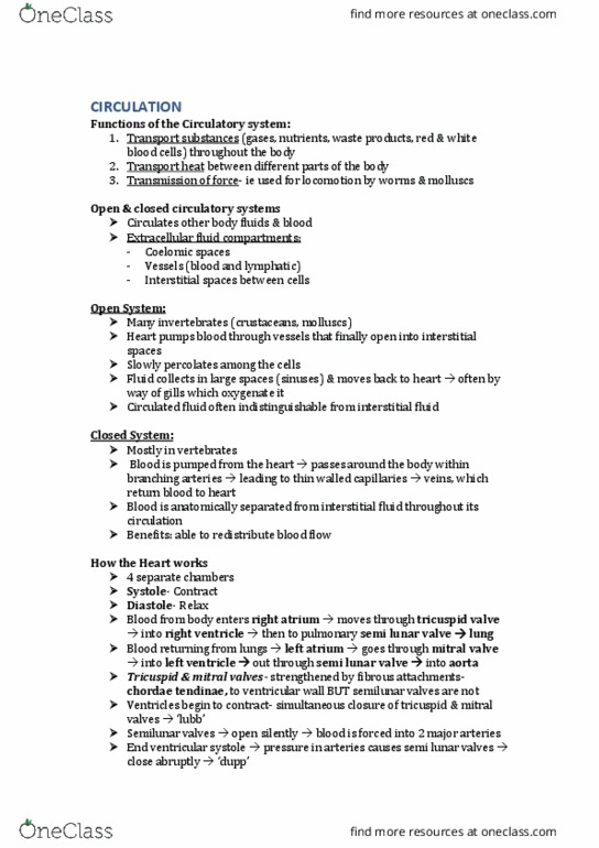
Closed
In some annelid worms and cephalopod molluscs
○
In all vertebrates
○
Heart pumps blood around body within branching arteries leading to
capillaries -> into veins that return blood to heart
○
Circulated fluid separated from interstitial
○
-
Features of hearts
Diastole: relaxation
Ventricles fill with blood (longer time)
○
Deoxygenated blood from superior vena cava -> enters right atrium (through
tricuspid valve) -> right ventricle -> pulmonary artery -> LUNGS
○
Oxygenated blood from pulmonary veins -> left atrium -> left ventricle ->
aorta -> BODY
○
-
Systole: contraction -> forcing blood out of heart
-
Source of contraction = myogenic - muscle in vertebrates - neurogenic -nerves in
invertebrates
-
Chordae tendineae: each flap of tricuspid valve is attached to strong strands of
connective tissue
-
Septum
-
Coronary arteries: openings in aorta - supply blood to heart muscle
-
Coronary sinus: veins that collect blood from heart muscle - delivers deoxygenated
blood to R atrium
-
Papillary muscles: contract to generate tension on chordae tendinae - prevents
backflow (in ventricles)
-
Septomarginal trabecula: moderator band - muscular band of heart tissue in R
ventricle - from base of papillary muscle - importance in electrical conduction
-
Arteriosclerosis/atherosclerosis: diseases of arteries which affect blood pressure + flow -
can cause heart attack and stroke
Cardiac muscle
Striated
-
Electrical depolarisation -> contraction
-
Muscle cells interconnected -> intercalated discs: microscopic features that mark
connection b/w 2 muscle cells - cells held together with gap junctions (electrical
connections) and desmosomes
-
Pacemaker = SA node
-
Describe sequence of electrical and mechanical events during cardiac cycle of mammal
SA node starts AP in right atrium
-
Atria contracts -> pushes blood into ventricles
-
AP triggers AV node -> 0.1 second delay (allows atrial contraction to finish before
ventricular contraction begins)
-
AP goes down atrioventricular bundle -> down interventricular septum -> apex of
heart
Rapid conduction down AV bundle and Purkinje fibres (fast conducting cells
that stimulate ventricles to contract - cells originate from AV bundle and
radiate throughout the ventricle muscle)
○
-
Ventricular contraction propagated from apex expels blood from heart -> systole
-
Distinguish b/w arteries, veins, and capillaries
Arteries
Blood away from heart
○
Elastic muscle walls - reduce fluctuations in blood pressure - Windkessel
vessels
○
Thick wall
○
Small lumen
○
-
Veins
Return blood to heart
○
Thin walls
○
Thick lumen
○
Thin layer of elastic
○
Capacitance vessels - most of total blood volume held in veins
○
-
Microcirculation
b/w arteries and veins
○
Arterioles
○
True capillaries: sites of exchange b/w blood and tissues
○
Venules
○
RBCs must pass through in single file
○
Wall = Single layer of cell
○
Very small lumen
○
Fluid that leaks out of capillaries -> transported back to heart via lymphatic
system
○
-
How are blood pressure and flow regulated in mammals?
In circulatory system as a whole
Regulated by negative feedback control
Baroreceptors (pressure)
Great veins□
Aortic arch□
Carotid sinus□
§
Chemoreceptors (chemicals)
Carotid body: O2□
Aortic body: CO2 and pH□
§
Integration in vasomotor and cardiac centres of brain
§
Sympathetic and parasympathetic outputs - control timing and strength
of heartbeat
§
○
Regulation affected by blood-borne hormones
○
-
Individual tissue level
Intrinsic mechanisms
Levels of metabolites (eg. Lactic acid, CO2, and ATP products)
§
○
Extrinsic mechanisms
Release of hormone noradrenaline - induces vasoconstriction of arterial
vessels, stimulated by neural input from higher centres
§
○
-
Response to exercise
Exercise -> decrease O2 and increase CO2 -> chemoreceptors (carotid and aortic bodies)
increased rate and strength of heartbeat -> increased gas exchange in lungs + increased
blood to muscles
vasoconstriction of some arteries -> increased blood to muscles
Exercise -> increased local metabolites -> local dilation of blood vessels -> increased blood
to muscles
Awareness in higher centres of brain -> vasodilation of arteries to muscle -> increased
blood to muscles
Increased blood to muscles - negative feedback on O2 and CO2 levels and metabolites
Fish circulation
4 chambered heart
-
Chambers fill and empty in sequence
-
Valves prevent reverse flow
-
Circulation
Wednesday, 30 May 2018
11:50 am

Why do animals have circulatory systems?
Transfer of O2 and nutrients
-
Waste products removed
-
Communication via hormones
-
Temp regulation + reproduction
-
Animals w/o rely on diffusion - small and thin
Jellyfish
○
Sponge
○
Flatworm
○
Corals and sea anemones
○
HIGH SA:VOLUME RATIO for diffusion
○
-
Animals w system transport by convection
Convection: bulk flow of a fluid (air or water)
Movement assisted by convection - faster than diffusion
§
○
-
Open vs. closed systems
Both contain pumps - heart
-
Open
In many invertebrates (eg. Crustaceans and some molluscs)
○
Heart pumps fluid through vessels that open into INTERSTITIAL SPACES
○
Fluid spreads among cells -> slowly returns to heart - often by gills that
oxygenate fluid on return
○
Cant distinguish circulated fluid from interstitial fluid
○
-
Closed
In some annelid worms and cephalopod molluscs
○
In all vertebrates
○
Heart pumps blood around body within branching arteries leading to
capillaries -> into veins that return blood to heart
○
Circulated fluid separated from interstitial
○
-
Features of hearts
Diastole: relaxation
Ventricles fill with blood (longer time)
○
Deoxygenated blood from superior vena cava -> enters right atrium (through
tricuspid valve) -> right ventricle -> pulmonary artery -> LUNGS
○
Oxygenated blood from pulmonary veins -> left atrium -> left ventricle ->
aorta -> BODY
○
-
Systole: contraction -> forcing blood out of heart
-
Source of contraction = myogenic - muscle in vertebrates - neurogenic -nerves in
invertebrates
-
Chordae tendineae: each flap of tricuspid valve is attached to strong strands of
connective tissue
-
Septum
-
Coronary arteries: openings in aorta - supply blood to heart muscle
-
Coronary sinus: veins that collect blood from heart muscle - delivers deoxygenated
blood to R atrium
-
Papillary muscles: contract to generate tension on chordae tendinae - prevents
backflow (in ventricles)
-
Septomarginal trabecula: moderator band - muscular band of heart tissue in R
ventricle - from base of papillary muscle - importance in electrical conduction
-
Arteriosclerosis/atherosclerosis: diseases of arteries which affect blood pressure + flow -
can cause heart attack and stroke
Cardiac muscle
Striated
-
Electrical depolarisation -> contraction
-
Muscle cells interconnected -> intercalated discs: microscopic features that mark
connection b/w 2 muscle cells - cells held together with gap junctions (electrical
connections) and desmosomes
-
Pacemaker = SA node
-
Describe sequence of electrical and mechanical events during cardiac cycle of mammal
SA node starts AP in right atrium
-
Atria contracts -> pushes blood into ventricles
-
AP triggers AV node -> 0.1 second delay (allows atrial contraction to finish before
ventricular contraction begins)
-
AP goes down atrioventricular bundle -> down interventricular septum -> apex of
heart
Rapid conduction down AV bundle and Purkinje fibres (fast conducting cells
that stimulate ventricles to contract - cells originate from AV bundle and
radiate throughout the ventricle muscle)
○
-
Ventricular contraction propagated from apex expels blood from heart -> systole
-
Distinguish b/w arteries, veins, and capillaries
Arteries
Blood away from heart
○
Elastic muscle walls - reduce fluctuations in blood pressure - Windkessel
vessels
○
Thick wall
○
Small lumen
○
-
Veins
Return blood to heart
○
Thin walls
○
Thick lumen
○
Thin layer of elastic
○
Capacitance vessels - most of total blood volume held in veins
○
-
Microcirculation
b/w arteries and veins
○
Arterioles
○
True capillaries: sites of exchange b/w blood and tissues
○
Venules
○
RBCs must pass through in single file
○
Wall = Single layer of cell
○
Very small lumen
○
Fluid that leaks out of capillaries -> transported back to heart via lymphatic
system
○
-
How are blood pressure and flow regulated in mammals?
In circulatory system as a whole
Regulated by negative feedback control
Baroreceptors (pressure)
Great veins□
Aortic arch□
Carotid sinus□
§
Chemoreceptors (chemicals)
Carotid body: O2□
Aortic body: CO2 and pH□
§
Integration in vasomotor and cardiac centres of brain
§
Sympathetic and parasympathetic outputs - control timing and strength
of heartbeat
§
○
Regulation affected by blood-borne hormones
○
-
Individual tissue level
Intrinsic mechanisms
Levels of metabolites (eg. Lactic acid, CO2, and ATP products)
§
○
Extrinsic mechanisms
Release of hormone noradrenaline - induces vasoconstriction of arterial
vessels, stimulated by neural input from higher centres
§
○
-
Response to exercise
Exercise -> decrease O2 and increase CO2 -> chemoreceptors (carotid and aortic bodies)
increased rate and strength of heartbeat -> increased gas exchange in lungs + increased
blood to muscles
vasoconstriction of some arteries -> increased blood to muscles
Exercise -> increased local metabolites -> local dilation of blood vessels -> increased blood
to muscles
Awareness in higher centres of brain -> vasodilation of arteries to muscle -> increased
blood to muscles
Increased blood to muscles - negative feedback on O2 and CO2 levels and metabolites
Fish circulation
4 chambered heart
-
Chambers fill and empty in sequence
-
Valves prevent reverse flow
-
Circulation
Wednesday, 30 May 2018 11:50 am
Document Summary
Animals w/o rely on diffusion - small and thin. Convection: bulk flow of a fluid (air or water) Movement assisted by convection - faster than diffusion. In many invertebrates (eg. crustaceans and some molluscs) Heart pumps fluid through vessels that open into interstitial spaces. Fluid spreads among cells -> slowly returns to heart - often by gills that oxygenate fluid on return. Heart pumps blood around body within branching arteries leading to capillaries -> into veins that return blood to heart. Deoxygenated blood from superior vena cava -> enters right atrium (through tricuspid valve) -> right ventricle -> pulmonary artery -> lungs. Oxygenated blood from pulmonary veins -> left atrium -> left ventricle -> aorta -> body. Systole: contraction -> forcing blood out of heart. Source of contraction = myogenic - muscle in vertebrates - neurogenic - nerves in invertebrates. Chordae tendineae: each flap of tricuspid valve is attached to strong strands of connective tissue.