BIOM20001 Lecture Notes - Lecture 5: Hydrophile, Fibronectin, Entropy


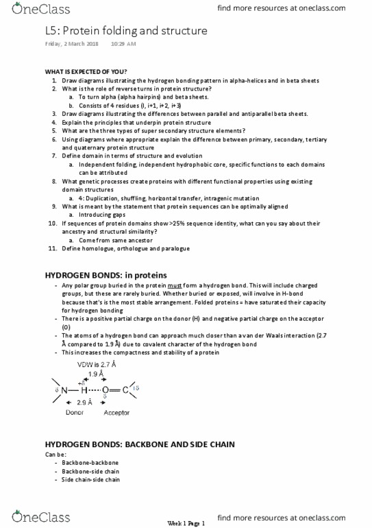
Any polar group buried in the protein must form a H-bond. This is because they MUST form
their lowest energy conformation. This will include charged groups, but these are rarely
buried.
•
+ve partial charge on the donor H, -ve partial charge on acceptor O
•
The atoms of a H-bond can approach much closer than a VDW interaction due to covalent
character of the H-bond. There is some sharing of electrons. This increases compactness and
stability of a protein by allowing residues to approach closer.
•
VDW: close packing of atoms
•
Can have backbone-backbone, backbone-side chain and side chain-side chain H bonds
•
Backbone-backbone H-bonds especially facilitates close packing of polypeptide
•
•
NH residue i to C=O residue i-4 (so NH bonds with residue backwards towards the
N terminus)
▪
3.6 residues per turn (100° per residue)
▪
Height of one turn is 5.4 Å (1.5 Å axial rise per residue)
▪
Side chains project outwards from helix axis
▪
Alpha-helix
○
Close packing of polypeptide backbone is achieved by several hydrogen bonding patterns
(secondary structure). Note: H-bonds should always be described donor to acceptor NH to
C=O. MUST SPECIFY WHICH IS DONOR GROUP AND WHICH IS ACCEPTOR. e.g. NH is donor and
C=O is acceptor
•
H-bonds in proteins
5-6 Proteins folding and structure
Friday, 28 February 2014
10:46 PM
MCB Page 1

▪
Right handed helix
▪
Abundant
▪
φ, ψ-57°, -47°
▪
All NH and C=O within the helix (except four at each end) form favourable internal
hydrogen bonds
▪
Peptide bond dipoles add together, giving a macrodipole
▪
▪
Alpha-helices often amphipathic, allowing burial of hydrophobic side chains by
packing 2 or more alpha-helices together (coiled-coil: alpha-helices have a heptad
repeat (7 residues labelled as abcdefg), creating a stripe of hydrophobic a and d
amino acids) or packing an alpha-helix against a beta-sheet
▪
Let's say we want to have an alpha coil where one side of is hydrophobic. We
construct the helix with polar and non-polar residues. Abcdefgabcdefgabcdefg.
We make every bolded residue a hydrophobic residue. Every other one is polar.
Note it is an alternating 3/4 repeating pattern since each turn is 3.6 residues. This
gives us a strip of hydrophobic side chains along one side of the helix. If we get
two helixes like that the hydrophobic stripes will stick together forming a
hydrophobic core.
▪
▪
Made of individual beta strands
▪
NH residue i to C=O residue j
▪
Alternating side chains project above and below sheet. If one points up, the
adjacent points down. If you wanted one side to be hydrophobic and one side
hydrophilic just alternate between hydrophobic and hydrophilic residues
▪
A beta-sheet consists of 2 or more strands. The strand is the element.
▪
Beta-sheet
○
MCB Page 2
Document Summary
Any polar group buried in the protein must form a h-bond. This is because they must form their lowest energy conformation. This will include charged groups, but these are rarely buried. +ve partial charge on the donor h, -ve partial charge on acceptor o. The atoms of a h-bond can approach much closer than a vdw interaction due to covalent character of the h-bond. This increases compactness and stability of a protein by allowing residues to approach closer. Can have backbone-backbone, backbone-side chain and side chain-side chain h bonds. Backbone-backbone h-bonds especially facilitates close packing of polypeptide. Close packing of polypeptide backbone is achieved by several hydrogen bonding patterns (secondary structure). Note: h-bonds should always be described donor to acceptor nh to. Must specify which is donor group and which is acceptor. e. g. nh is donor and. Nh residue i to c=o residue i-4 (so nh bonds with residue backwards towards the.