BIOM20002 Lecture Notes - Lecture 34: Anterior Interventricular Sulcus, Coronary Sinus, Interventricular Septum

5 Sep 2018
School
Department
Course
Professor
Document Summary
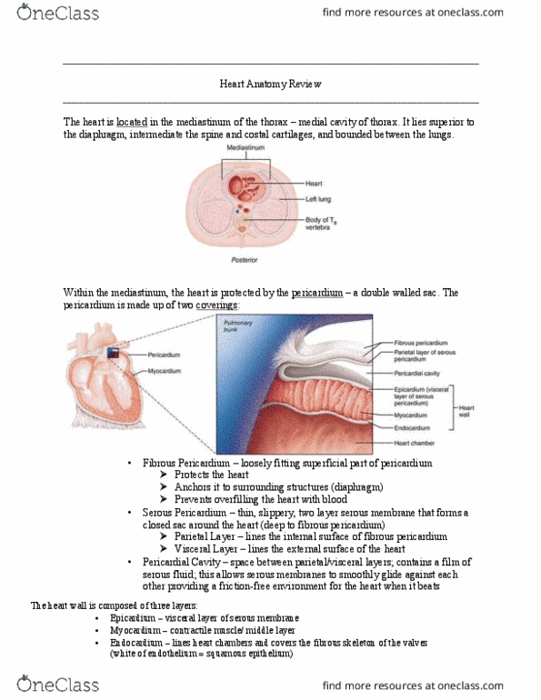
Lined internally both the surface of heart and lining of pericardium based on the how is it derived from development. Whenever the organ is expandable, it has a thin layer of serous membrane surrounding it. And also the wall of the cavity outside that organ has the same thin layer of serous membrane: while those organs expand and contract, they slip against their wall cavity in a smooth friction-free fashion, produce serous fluid. Potential space (little gap) between fibrous membrane and serous membrane containing serous fluid allowing friction free glide. Atria lie to the right, ventricles to the left. From left to right: right atrium coronary sulcus right ventricle anterior interventricular sulcus. Base where the vessels come out. There are little ear-like extensions of both atria atrial appendages. Left ventricle on the left border of the posterior view. See a bit of right atrium on the right border of the heart.