PSYC20006 Lecture Notes - Lecture 7: Compass, Rubik'S Cube, 8 Seconds

10 May 2018
School
Department
Course
Professor

Lecture 7
- One of the holy grails of bio psych has been to image the functioning of human
brain: wanted to know which bits of the brain you were using when; when a subject
performs a particular task (like playing tennis or recognising his friend from a crowd
or performing a visual search experiment), which bits of the brain are involved in
performing that task → fMRI can answer this question
- In the past, this question was answered using a technique called Positron Emission
Tomography (PET): involves administering subjects radioactive oxygen while they
perform 3450-=\a task (e.g. oxygen-15, exposing patient to significant amount of
ionizing radiation), brain areas more activated by the task which are responsible for
allowing that person to perform the task will absorb more radioactive oxygen (brain
areas that work harder will absorb more radioactive oxygen and become more
radioactive) → scan a person’s brain to find out which bits are the most radioactive,
how you determine which bits were most active while they were performing the task;
levels of radiation are low and within safe limits
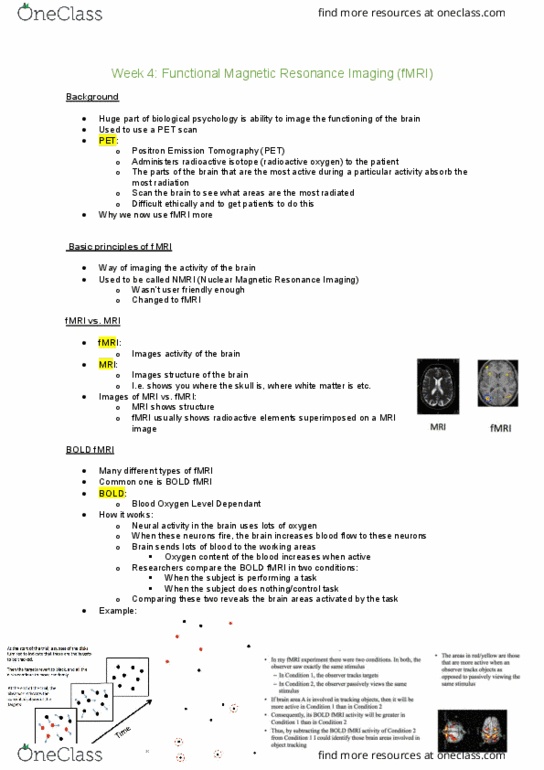
- fMRI: is a way of imaging the activity in the human brain (activity not structure);
originally called NMRI (Nuclear Magnetic Resonance Imaging) but the term nuclear
was thought to have too many negative connotations; functional sounded more user
friendly
-
-
- fMRI: measures activity of human brain
- MRI: measures structure of human brain
- Grey background showing structure is MRI, onto which fMRI (yellow bits) have been
superimposed; usually when you see fMRI you’re seeing fMRI superimposed onto
MRI
- Different types of fMRI
find more resources at oneclass.com
find more resources at oneclass.com

- BOLD fMRI: Blood Oxygen Level-Dependent
- Neural activity uses oxygen; when neurons fire APs (they use up oxygen) → brain
increases blood flow to them because they need more oxygen
- Because brain sends so much blood to active area, the oxygen content of blood
actually increases (brain overcompensates, anti-intuitive)
- Blood oxygen levels increase around neurons that are working harder
-
- BOLD fMRI: compare BOLD fMRI signals coming from the brain in two situations:
- 1) when the subject performs the task that you’re interested in
- 2) when the subject either does nothing or performs a control task
- Subtracting 2) from 1) reveals those areas of the brain that were preferentially
activated by the task of interest
-
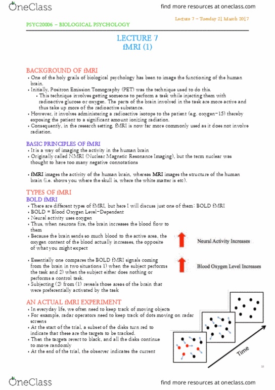
- Keep track of moving objects in our environment; e.g. when driving car need to keep
track of other road users so that we don’t run into them; radar operator need to keep
track of objects on our radar screens
find more resources at oneclass.com
find more resources at oneclass.com

-
- Subjects had to mentally keep track of targets which were the same colour as the
distractors
-
- Tracking only occurs in Condition 1
- Can identify the brain areas that were more active when observer was tracking
objects than when he was just passively viewing the objects
- Standard format for all fMRI
find more resources at oneclass.com
find more resources at oneclass.com
Document Summary
In the past, this question was answered using a technique called positron emission. Grey background showing structure is mri, onto which fmri (yellow bits) have been superimposed; usually when you see fmri you"re seeing fmri superimposed onto. Neural activity uses oxygen; when neurons fire aps (they use up oxygen) brain increases blood flow to them because they need more oxygen. Because brain sends so much blood to active area, the oxygen content of blood actually increases (brain overcompensates, anti-intuitive) Blood oxygen levels increase around neurons that are working harder. Bold fmri: compare bold fmri signals coming from the brain in two situations: 1) when the subject performs the task that you"re interested in. 2) when the subject either does nothing or performs a control task. Subtracting 2) from 1) reveals those areas of the brain that were preferentially activated by the task of interest. Subjects had to mentally keep track of targets which were the same colour as the distractors.