BMS1052 Lecture Notes - Lecture 17: Plantar Reflex, Cell Nucleus, Deep Brain Stimulation

30 May 2018
School
Department
Course
Professor

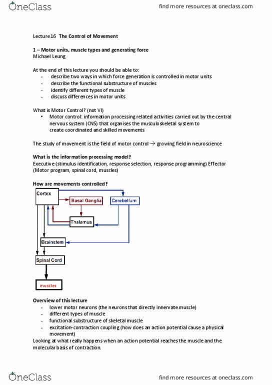
Week 7. Control of movement 3, 4 & 5
CONTROL OF MOVEMENT 3 – SPINAL REFLEXES AND PROPRIOCEPTION
• Topographical organisation of motor neurons:
o Proximal limb muscles – medial
o Distal limb muscles – lateral
o 1. Flexor-extensor rule: motor neurons that innervate flexor muscles are located
posteriorly to motor neurons that innervate extensor muscles.
o 2. Proximal-distal rule: motor neurons that innervate distal muscles (e.g., hand muscles)
are located lateral to motor neurons that innervate proximal muscles (e.g., trunk
muscles).
o This helps with functional organisation
• Renshaw cells –recurrent inhibition of motor neurons
o Example of a local circuit in the spinal cord
o Simplest circuit of only 2 neurons
o Function is not precisely known
o Each Renshaw cell synapses on dendrites f 1 or more alpha motor neurons and primary
input is from 1 or more motor neurons
o Inhibitory interneuron -> releases GABA -> suppresses further activation of the motor
neuron -> negative feedback loop with lower motor neuron
o May help reduce noise (prevent contraction in response to weak inputs)
o May help prevent muscular damage from over-excitation
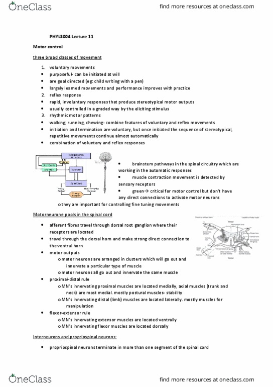
• There are two sensory receptors in the muscles:
-know where limbs are in space; helps maintain muscle length and tension, preventing muscle
overload and compensating for fatigue
1. Muscle spindles:
Stretch (length) receptors
In parallel with muscle fibres
2. Golgi tendon organs:
Tension (force) receptors
In series with muscle fibres
• Muscle spindles:
o Spindle receives distinct sensory and motor innervation
o In parallel with Extrafusal muscle fibres
o Fibrous capsule (fluid filled) of muscle spindle contains specialised intrafusal fibres
o Activity in muscle spindles can be affected in 2 ways:
1. Stretch of muscle
2. Contraction of Intrafusal fibres
find more resources at oneclass.com
find more resources at oneclass.com

o Intrafusal vs Extrafusal:
Extrafusal and Intrafusal are innervated by Ia sensory neurons
-they have stretch sensitive ion channels to signal muscle length
Distinguishing feature - cell nuclei are clustered in the center of the fibre.
Intrafusal
Extrafusal
o Intrafusal fibres : responsible for
changing spindle length by contracting
o in spindle
o striated muscle and have sarcomeres
as seen in standard skeletal muscle
o There are 3 types that all detect
muscle length:
1. Nuclear chain
2. Nuclear bag I – also responds to
sudden changes in length
3. Nuclear bag II
o Nucleated region of Intrafusal is more
streth -> where primary afferents
terminate so more sensitive to length
changes
o Extrafusal fibres are responsible for
generating force by shortening
o Outside spindle
o Targeted by alpha motor neurons to
shorten bulk of muscle
o Sensory afferents:
Ia
II
o Spiral winding around nuclear region
o Contacts all Intrafusal fibres
o High conduction velocity
o Firing rate is highest when muscle is
stretched but largest firing rate occurs
during rapid stretch
o Larger and more heavily myelinated ->
enables rapid sensation and short
reflexes
o Primary afferent -> can signal small
changes in length
o Encode muscle length and rate of length
change
o Contacts bag 2 and chain fibres
o Slower conduction velocity
-> more sluggish response -> responds
slowly to rapid changes
o More lateral -> termination to side of
primary endings
o Axons have slower conduction velocity
o Ol eode positio do’t respod to
brief taps or vibrations)
o Encodes only muscle length
o Group II - secondary endings in spindles
& mechanoreceptors – 30-60 m/s
find more resources at oneclass.com
find more resources at oneclass.com

o Group Ia - primary endings in spindles –
60-120 m/s
o Group Ib - from tendon organs – 60-120
m/s
o Have large diameters
o What affects conduction velocity?
Dimeter, myelination, temperature, damage
• Gamma co-activation – maintaining sensitivity of the spindle
o Maintains sensitivity to small changes, important as after contraction of Extrafusal fibres,
muscles spindle is slack -> lose its ability to signal muscle length (sensory neuron goes
offlie ad lose sustaied firig rate
o Gamma motor neurons target Intrafusal fibres to shorten the muscle spindle
o Opposing affects:
Alpha activation decreases Ia activity
Gamma activation increases Ia activity
o By continually adjusting length of spindle -> sensitivity is increased
• Myotatic reflex (aka tendon reflex – tapping on tendon)
o most basic reflex
o does not involve golgi tendon
o group Ia sensory neurons synapse on alpha motor neurons and interneurons
o a monosynaptic feedback loop mediates the myotatic reflex
o Tendon jerk is largest when muscle length is optimal length for contraction. If muscle is
short, tedo is slak. If usle is log, spidles hae high akgroud atiit ad do’t
signal additional stretch
o Lower motor neurons also have input from cortex -> descending inhibition -> modulates
size of reflexive movement
o Jendrassik manouever: shows that reflexes are modulated by descending inhibition
-> when clasping hands, reduces inhibition -> reflex in leg gets bigger (form of distraction)
find more resources at oneclass.com
find more resources at oneclass.com
Document Summary
Control of movement 3 spinal reflexes and proprioception: topographical organisation of motor neurons, proximal limb muscles medial, distal limb muscles lateral, 1. Flexor-extensor rule: motor neurons that innervate flexor muscles are located posteriorly to motor neurons that innervate extensor muscles: 2. Know where limbs are in space; helps maintain muscle length and tension, preventing muscle overload and compensating for fatigue: muscle spindles: In parallel with muscle fibres: golgi tendon organs: In series with muscle fibres: muscle spindles, spindle receives distinct sensory and motor innervation. In parallel with extrafusal muscle fibres: fibrous capsule (fluid filled) of muscle spindle contains specialised intrafusal fibres, activity in muscle spindles can be affected in 2 ways, stretch of muscle, contraction of intrafusal fibres. Extrafusal and intrafusal are innervated by ia sensory neurons. They have stretch sensitive ion channels to signal muscle length. Distinguishing feature - cell nuclei are clustered in the center of the fibre.