BMS1052 Lecture Notes - Lecture 12: Snellen Chart, Ciliary Muscle, Visual Acuity

18 Aug 2018
School
Department
Course
Professor
Document Summary
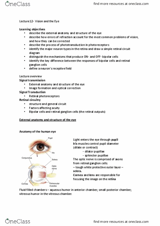
Lecture 12 : vision and the eye i. Anatomy of the human eye the eye is a fluid filled globe protected by a tough, white outer layer called the sclera. Light enters the pupil ( a transparent membrane) iris is the coloured region surrounding the pupil iris muscles control the pupil diameter called dilator pupillae. Sphincter pupillae the output of the eye comprises of the retinal ganglion cells that exit via the optic nerve. Within the fluid filled globe, aqueous humour is in the anterior chamber ( front chamber),while vitreous humour is in the vitreous chamber. The small posterior chamber lies between the anterior chamber and the vitreous chamber. Note : retinal ganglion cells are considered a. The image here is the back of the eye. Optic disk or blind spot - has no photoreceptors, is the site of blood vessel entry and exit and optic nerve entry and exit.