BMS2011 Lecture Notes - Lecture 4: Muscular System, Synovial Joint, Medullary Cavity

30 May 2018
School
Department
Course
Professor

Week 2. Musculoskeletal and Axial
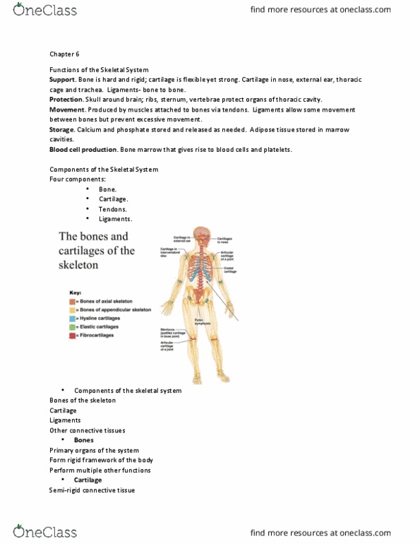
Musculoskeletal system
MUSCULOSKELETAL SYSTEM
• Skeletal tissues and muscles are derived from mesoderm
Paraxial -> somite -> dermatome (dermis/epidermis), myotome (muscle), sclerotome
(bone/cartilage)
• Both blood and bone are connective tissue, what differentiates them is composition of
extracellular matrix
• Bone: tissue composition:
o 1. Fibres (organic): collagen, provides tensile strength
o 2. Ground substance (extracellular matrix: inorganic): calcium phosphate, hydroxide and
carbonate -> forms hydroxyapatite crystals (mineral components), most of compressive
strength
o Osteoblasts: cells that produce extracellular matrix (osteoid), found on surface of any
developing bone area
-original cell, creates bone
o Osteocytes: osteoblasts that have become surrounded by matrix, lie in space within matrix
called lacunae (maintain bone matrix)
-mature cell surrounded by osteoid
o Osteoclasts: capable of degrading and reabsorbing matrix, found on surface of any bone
undergoing remodelling
-destroys bone
• Bones are alive, receive nutrients through diffusion
find more resources at oneclass.com
find more resources at oneclass.com

• Bone tissue organisation:
Compact (Lamellar) bone
o Osteon (haversian system) consists of:
o Central haversian canal and lamellae (concentric rings)
-blood vessels and nerves inside (blood supply for cells)
-osteocytes between lamellae
o Lacunae: spaces between lamellae containing osteocytes
-like a lake
o Canaliculi: canals radiating from lacunae
-channels between adjacent cells
o Perforatig Volka’s caals: join osteon central
canals (perpendicular)
Spongy (Trabecullar) bone
o Lamellae form trabeculae (branching plates) not osteons
-no canals/haversian
o Nutrients via diffusion along canaliculi from the endosteum
o Red bone marrow in proximal humerus and femur, vertebrae,
sternum, ribs and pelvis (used as nursery for differentiation of cells
eg. RBC, WBC)
o Organised to minimise stress and strain
(there are internal pathways that follow the way that loads are
being born in the joints)
find more resources at oneclass.com
find more resources at oneclass.com

• Bone tissue variation: classification
Long
o Elongated shaft and enlarged end
o Eg. humerus, femur, tibia
o Landmarks and regions:
o Diaphysis: cylinder of compact bone (shaft) with medullary cavity (yellow
marrow, fat -> makes it long)
-hollow inside
o Metaphysis: between diaphysis and epiphysis
-band of cartilage serving as growth plate
-stays open during growth
o Epiphysis: enlarged end with compact and trabeculae bone: supports
articular cartilage
-end of bone, smooth appearance
o When diaphysis and epiphysis connect -> stop growing
Irregular
o Complex shape
o Eg. vertebrae and some skull bones
Short
o Geometrically equivalent in all directions
o Eg. carpals and tarsals
Flat
o Parallel layers of compact bone with trabecular
o Eg. cranium, sternum, ribs, scapula
Sesamoid
o Develop in tendons
o Eg. patella
find more resources at oneclass.com
find more resources at oneclass.com
Document Summary
Musculoskeletal system: skeletal tissues and muscles are derived from mesoderm. Paraxial -> somite -> dermatome (dermis/epidermis), myotome (muscle), sclerotome (bone/cartilage: both blood and bone are connective tissue, what differentiates them is composition of extracellular matrix, bone: tissue composition, 1. Fibres (organic): collagen, provides tensile strength: 2. Ground substance (extracellular matrix: inorganic): calcium phosphate, hydroxide and carbonate -> forms hydroxyapatite crystals (mineral components), most of compressive strength: osteoblasts: cells that produce extracellular matrix (osteoid), found on surface of any developing bone area. Original cell, creates bone: osteocytes: osteoblasts that have become surrounded by matrix, lie in space within matrix called lacunae (maintain bone matrix) Mature cell surrounded by osteoid: osteoclasts: capable of degrading and reabsorbing matrix, found on surface of any bone undergoing remodelling. Destroys bone: bones are alive, receive nutrients through diffusion, bone tissue organisation: Compact (lamellar) bone: osteon (haversian system) consists of, central haversian canal and lamellae (concentric rings) Blood vessels and nerves inside (blood supply for cells)