BIO282 Lecture Notes - Lecture 5: Agarose Gel Electrophoresis, Ethidium Bromide, Agarose


DNA Detection
• Gel electrophoresis
• Southern Blotting
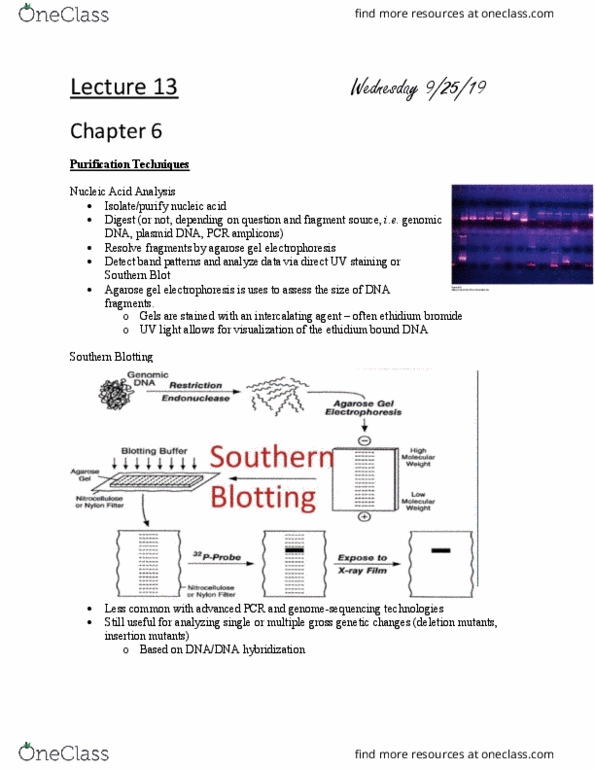
Agarose Gel Electrophoresis
• Agarose gel electrophoresis is a routine techniques used to separate nucleic acid (DNA
and RNA) molecules.
• Agarose is a complex network of polysaccharide molecules prepared from seaweed;
forms a series of pores in the gel form.
• DNA molecules migrate through these pores when an electric current is applied to the gel.
DNA is negatively charged, it moves towards the +vepole. Smaller fragments run faster
• DNA is visualised by staining with ethidium bromide, or other commercially available
stains, that forms a complex with DNA. Bromide binds to DNA so it is not safe and
alternatives are being sought.
• The gel is placed on a UV light to visualize the DNA bands.
• Analysis of DNA fragments in a gel can determine both the quality and quantity of DNA
present in a sample. You can tell how much DNA there is and how intact or broken it
is.
The sample is put into little dents called wells in the plate and stained.
The bands are the DNA molecules. Smaller fragments run faster and will be further from
the well. The biggest will move the least from the well.
Kb is kilo bases (not kilo bytes)

Example under UV
Most of the DNA should be in supercoiled but because of impurities, you will mostly see
relaxed. If you run three different examples you will see three different bands.
• Supercoiled state
• Relaxed circle
• Linear form
The suer coiled moves fastest because it is tightly packed and the linear form is the
slowest and its length takes more time to move.
Locating DNA Fragments with Southern Blotting and Probes
It is called southern blotting because it is blotting the DNA much like blotting paper in the
past blotted up excess ink from pens. Southern is from the surname from the scientist
that developed it.
Probe: DNA or RNA with a base sequence complementary to a sequence in the gene of
interest

Southern Blotting and Probes
Southern blotting, developed by E. M. Southern in 1975, is used to reveal identity, abundance
and size of a specific DNA.
• Some of the applications of Sothern blotting include
o identification of -
• a gene and gene mutations
• the number of copies of a particular gene
• related genes in different organisms
• genetic diseases
• tests in forensic science as
• Paternity testing
• Personal identification
• Sex determination
• in food industry
• confirmation of biological ingredients
When genomic DNA is cut by a restriction enzyme it generates tens of thousands of DNA pieces
that appear as a smear on an agarose gel (2)
What is the purpose of Southern Blotting?
It is to separate it from the paper and to find a specific gene. The complimentary DNA is
found by the probe. It is detected through the radioactivity of the probe seen with x ray.
This has been a common method but now there are concerns about safety and other
methods were developed. The probe is still the same but instead of radioactivity they
use biotin.
The Southern Hybridization technique can identify a specific piece of DNA or a gene with the
help of a probe.
Probe is a sequence of nucleotides with a base sequence complementary to the target gene. A
probe bound to the target DNA is identified by using one of the several detection systems
available (1)
Document Summary
Dna is negatively charged, it moves towards the +vepole. Smaller fragments run faster: dna is visualised by staining with ethidium bromide, or other commercially available stains, that forms a complex with dna. You can tell how much dna there is and how intact or broken it is. The sample is put into little dents called wells in the plate and stained. Smaller fragments run faster and will be further from the well. The biggest will move the least from the well. Most of the dna should be in supercoiled but because of impurities, you will mostly see relaxed. If you run three different examples you will see three different bands: supercoiled state, relaxed circle, linear form. The suer coiled moves fastest because it is tightly packed and the linear form is the slowest and its length takes more time to move. Locating dna fragments with southern blotting and probes.