BIOM3002 Lecture Notes - Lecture 3: Yolk Sac, Amniotic Sac, Umbilical Cord

26 May 2018
School
Department
Course
Professor

EMBYROLOGY LECTURE THREE
Gastrointestinal embryology:
• Lining of the abdominal cavity
• Gut formation
• Embryological defects
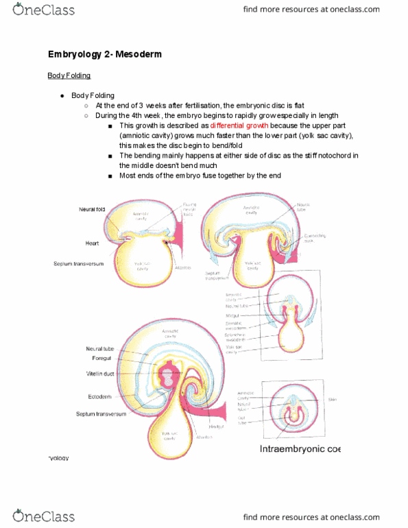
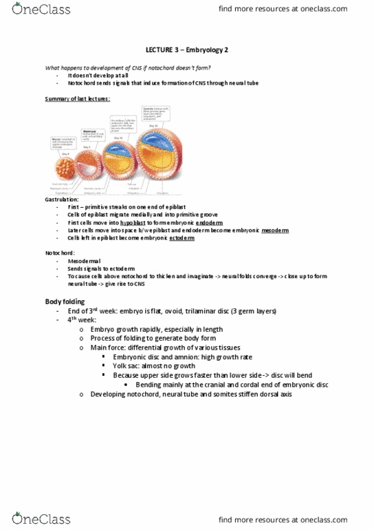
REVISION: WEEK 4 Embryonic Folding
• Embryo begins to fold [bend] along two axes: anterior–
posterior and medio-lateral
• 4 x folds: head, tail and 2 x lateral folds created by
amniotic cavity [lined with ectoderm]
Folding involves:
• Increased growth on dorsal surface of embryo proper
[increased neuroepithelium cf. notochord]
• Increased growth of amniotic sac and mesoderm layers at
margins
Yolk sac "squeezed", part of it excluded [outside embryo],
part inside embryo
Amniotic sac = ectoderm lined
Yolk sac = endoderm lined
Forms anterior-posterior tube and rest emerges out of
umbilicus
Note: embryo is floating in amniotic sac
Problems can emerge with failure embryonic folding, e.g.
abdominal wall defects at umbilical region
Connecting stalk = future umbilical cord
EXPLANATION:
1. From 4th week development = rapid development size and
shape
2. Trilaminar disc undergoes a process called embryonic folding
to create a basic 3D human body plan
3. Flat trilaminar disc -> cylinder [embryonic folding]
• The result of different rates of growth of embryonic
structures
• Occurs simultaneously in 2 planes: horizontal and median
4. Horizontal plane = 2 lateral body folds
5. Median plane= cranial and caudal fold
6. Cylinder 3 x layers: endoderm, mesoderm, ectoderm
find more resources at oneclass.com
find more resources at oneclass.com
Document Summary
Gastrointestinal embryology: lining of the abdominal cavity, gut formation, embryological defects. Revision: week 4 embryonic folding: embryo begins to fold [bend] along two axes: anterior , 4 x folds: head, tail and 2 x lateral folds created by posterior and medio-lateral amniotic cavity [lined with ectoderm] Folding involves: increased growth on dorsal surface of embryo proper. [increased neuroepithelium cf. notochord: increased growth of amniotic sac and mesoderm layers at margins. Yolk sac squeezed, part of it excluded [outside embryo], part inside embryo. Forms anterior-posterior tube and rest emerges out of umbilicus. Problems can emerge with failure embryonic folding, e. g. abdominal wall defects at umbilical region. Ruptures to become mouth at end 4th week development. Midgut: remains connected to yolk sac until 5th week development. Ruptures to form anal and urogenital openings at 7th week development. Chorionic cavity progressively disappears: filled with fluid which progressively decreases until 12 weeks when it is obliterated.