BIOL1003 Lecture Notes - Lecture 4: Hyaline Cartilage, Synovial Joint, Synovial Fluid

19 May 2018
School
Department
Course
Professor

Describe the joints of the axial and appendicular skeleton and the structure of synovial joints
50% bulk of the human body is muscle
-
Mostly skeletal muscle working in conjunction with skeleton to produce movement and locomotion
-
Skeletal muscles arranges in complex sets, antagonistic pairs
-
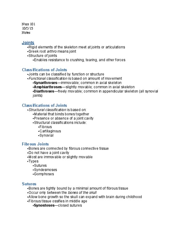
Joint/articulation - where 2 bones come together
-
Types of joints
Joint
Description
Example
Fibrous joints
Fixed/immovable joint
-
Bones held together by
connective tissues
-
Bones connected to form rigid,
supporting structures
-
Consist of 2 bones that are
united by fibrous tissues
-
Found where important to have
joints which exhibit little or no
movement
-
Sutures - fibrous joints between bones of the skull
-
Bones of skull
-
Teeth and jaws - gomophosis = joint between tooth and its socket (consists
of pegs fitted into sockets and held in place by ligament)
-
Jaw
-
Cartilaginous joints
Unites 2 bones by cartilage
-
Slight/limited movement
-
Bones joined by interposed
cartilage fasted to bones by
connective tissues
-
Cartilage of some joints where
much strain is placed, may be
reinforced by additional collagen
fibres called FIBROCARTILAGE
-
Between adjacent vertebra
-
Between coxal bones
-
Between ribs and sternum
-
Cartilage in the epiphyseal plates of growing long bones
-
Synovial joints
Freely moving
-
Bound together by ligaments
and connective tissue forming
fluid filled joint cavity
-
In cavity ends of bones covered
in articular cartilage, provides
smooth surface where bones
meet
-
Outer articular capsule helps
maintain bone alignment while
elastic synovial membrane
secretes and contain lubricating
synovial fluid
-
Like when cut open lamb shank
-
Joint cavity -filled with synovial
fluid
-
Cavity enclosed by a joint
capsule which holds bones
together and allows for
movement
-
Ligaments and tendons outside
joint capsule contribute to
strength of joint
-
Synovial membrane lines joint
cavity everywhere except over
articular cartilage
-
Synovial fluid produced by
membrane - complex mixture of
polysaccharides, proteins, lipids
and cells
-
Elbow
-
Shoulder
-
Hip
-
Knee
-
Most in appendicular skeleton
-
Screen clipping taken: 30/03/2017 9:58 PM
4a)
Thursday, 30 March 2017
1:14 PM
4. Muscles and Joints Page 1

and cells
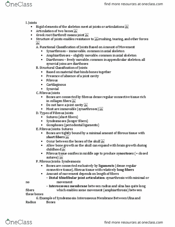
Types of synovial joints
Type
Description
Example
Hinge joint
Allows movement in one plane only
-
Extension and flexion
-
Elbow joint - humerus, ulna, radius
-
Articulation of atlas and skull (occipital
condyles) - allows nodding of head
-
Knee - femur, tibia
-
Ankle - talus, tibia, fibula
-
Interphalangeal - between phalanges of
fingers or toes
-
Pivot joint
Allows movement around one axis only ie. Rotary movement
Articulation between atlas and axis in
cervical vertebra
-
Allows no movement of head ie. rotation
-
Ligaments and muscles of neck limit this
movement
-
Between head of radius and proximal shaft
of ulna in the arm
-
Distal end of radius and ulna
-
Gliding or
plane joint
The articular surfaces glide over one another
Intercarpal joints formed between carpal
and bones of the write
-
Intertarsal and intermetatarsal joints
-
Sterno-clavicular joint - clavicle and
manubrium of sternum
-
Ball and socket
joint
The swollen rounded end of one bone fits into a cup-shaped
cavity of another, and allows movement in all planes
Only 2 in whole body
Shoulder joint - humerus and scapula
Hip joint - coxal bone and femur
Bones in synovial joints held together by the articular capsule and supporting ligaments
4. Muscles and Joints Page 2

Explain how muscle action causes movement at the joints and define an antagonistic muscle pair
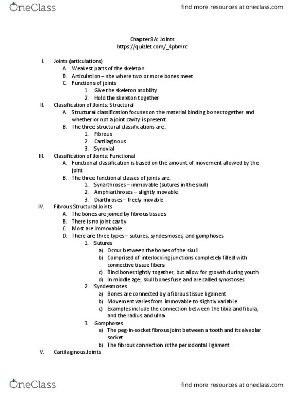
Movement about joints
ANGULAR movements - increase or decrease the angle between 2 adjoining bones in a single plane
Flexion
Bending of a joint
-
Usually reducing angle that 2 movable articulated bones make with each other
-
e.g. elbow, knee, neck (atlas and skull)
-
Screen clipping taken: 30/03/2017 10:07 PM
4b)
Thursday, 30 March 2017
1:19 PM
4. Muscles and Joints Page 3
Document Summary
Describe the joints of the axial and appendicular skeleton and the structure of synovial joints. 50% bulk of the human body is muscle. Mostly skeletal muscle working in conjunction with skeleton to produce movement and locomotion. Skeletal muscles arranges in complex sets, antagonistic pairs. Consist of 2 bones that are united by fibrous tissues. Found where important to have joints which exhibit little or no movement. Bones joined by interposed cartilage fasted to bones by connective tissues. Cartilage of some joints where much strain is placed, may be reinforced by additional collagen fibres called fibrocartilage. Bound together by ligaments and connective tissue forming fluid filled joint cavity. In cavity ends of bones covered in articular cartilage, provides smooth surface where bones meet. Outer articular capsule helps maintain bone alignment while elastic synovial membrane secretes and contain lubricating synovial fluid. Cavity enclosed by a joint capsule which holds bones together and allows for movement.