CHEM1112 Lecture Notes - Lecture 3: Electromagnetic Spectrum, Mass Spectrum, Chemical Formula

16 May 2018
School
Department
Course
Professor

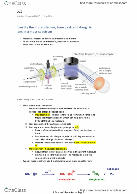
• Recognise that a fragmentation pattern in a mass spectrum can lead to structural information
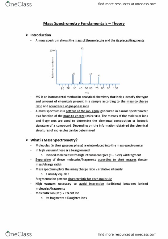
• Empirical formula= ratios- could be any structure- mass = molecular formula- mass spectrometry.
• Measure mass of molecules- ionised in vacuum, magnetic field, causing bend-circular paths- radius
dependent on m and z> some fragment into smaller ions (daughter ions)- help determine
structure of molecule > accelerated through electric field> how far ions moved= radius= mass
• Ions separated according to m/z (mass/charge)
• Molecular ion: heaviest ion, from loss of one electron from parent molecule, mass molecular ion=
mass parent ion
• Base peak: most intense signal, intensity set to 100%
Electrospray ionisation/Electrospray mass spec: aerosol, mild acid/buffer- ions hydrogenised, gentle-can
analyse proteins.
Involves absorbtion of part of electromagnetic spectrum- triggers change of energy level occupancy in
molecule, energy absorbed= signal in spectrum= structural info.
• Recognise the isotope distribution characteristic of bromine and chlorine containing compounds
in the mass spectrum
Each ion in mass spec registers specific isotopes of elements present- some isotopes more common, but
some exist in ~~equal amounts, ie: 2 molecular ion peaks.
Bromine79= 51%, Br81= 49%
Cl35= 75%, Cl37= 25%
High resolution mass spec: uses most abundant isotope.
• Use the information that IR and UV-visible spectra provide aid in the determination of
structures
• Identify the number and type of hydrogen environments in a molecule and predict the number
of signals in a 1H NMR spectrum
Nuclear magnetic Resonance: radio frequency region, flipping spin of nucleus in a magnetic field- used in
NMR & MRI.
Many nuclei are NMR active (H, C13, P31, F19)- resonate at particular frequency in magnetic field.
Nuclear spin = I= +/- 1/2. if I = 0 = inactive, if I > 1/2 = complex.
find more resources at oneclass.com
find more resources at oneclass.com
Document Summary
Ions separated according to m/z (mass/charge: molecular ion: heaviest ion, from loss of one electron from parent molecule, mass molecular ion= mass parent ion, base peak: most intense signal, intensity set to 100% Electrospray ionisation/electrospray mass spec: aerosol, mild acid/buffer- ions hydrogenised, gentle-can analyse proteins. Involves absorbtion of part of electromagnetic spectrum- triggers change of energy level occupancy in molecule, energy absorbed= signal in spectrum= structural info: recognise the isotope distribution characteristic of bromine and chlorine containing compounds in the mass spectrum. Each ion in mass spec registers specific isotopes of elements present- some isotopes more common, but some exist in ~~equal amounts, ie: 2 molecular ion peaks. High resolution mass spec: uses most abundant isotope: use the information that ir and uv-visible spectra provide aid in the determination of structures. Identify the number and type of hydrogen environments in a molecule and predict the number of signals in a 1h nmr spectrum.