CAM101 Lecture Notes - Lecture 15: Basal Lamina, Loose Connective Tissue, Basal Body


Learning Objectives
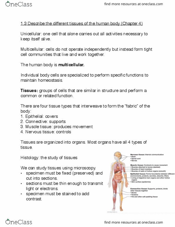
• Illustrate the two ways in which cells can form a tissue
• Describe the basic characteristics of epithelia, and be able to compare and contrast these with
the characteristics of the other three adult primary tissue types
• Demonstrate an understanding of the classification of epithelia
• Describe the structure, function and location of the various types of epithelia commonly found
in the human body
• Describe the structure and function of the five different types of cell junctions
• Compare and contrast the structure and function of microvilli, stereocilia and cilia
• Briefly describe the structure and function of the basement membrane
Key Features
1. Covers the body surfaces (internally and externally)
2. Boundary or Interface between different environments
• Protection, absorption, filtration, excretion, secretion, sensory reception
3. Forms almost all glands of the body
Characteristics
1. Cellularity
• Virtually no extracellular material between cells
2. Cell Junctions
• Numerous cell junctions to help them form a tissue
3. Basal Lamina
• Every epithelium sits upon a basal lamina, separates epithelium from connective tissue
• Basal lamina combines with a layer formed by connective tissue called the reticular
lamina to form a basement membrane.
4. Polarity
• Able to recognise what surface is their top (apical), what surfaces are to their sides
(lateral), and what surface is the bottom (basal)
5. Supported
• Loose connective tissue provides structural and metabolic support to overlying epithelial
tissue
6. Avascular (Lacks blood vessels)
• Epithelial tissue rely upon loose connective tissue to receive nutrients and remove
wastes.
7. Regeneration
• Contain stem cells that can undergo mitosis to produce new epithelial cells, important in
relation to healing.
8. Cell Membrane Specialisations
• Apically: Microvilli, cilia and/or stereocilia may be present
• Laterally: Cell Junctions exist
• Basally: Cell Junctions and basal lamina are present
Classification
1. Number of Cell Layers
• Simple Epithelium: One cell layer thick (all cells touch basal lamina)
• Usually specialised as lining of vessels and cavities, where they regulate passage of
substances into the underlying tissue
• Stratified Epithelium: >1 cell layer thick (not all cells touch basal lamina)
find more resources at oneclass.com
find more resources at oneclass.com

• Generally serve to protect and in some cases secrete keratin and prevent water
loss
• Pseudostratified Epithelium: Appears stratified but is really one cell layer thick (all cells
touch basal lamina)
2. Shape of the Cells on the Surface
• Squamous: Elongated and thin
• Cuboidal: Cube-shaped
• Columnar: Tall and thin
Common Epithelial
Simple Squamous
Barrier of least resistance to diffusion, while also providing a smooth frictionless surface
• Line surfaces where diffusion of gases/liquids occur.
• Covers many organs
• Lines cardiovascular system (known as endothelium in blood vessels)
Simple Cuboidal
Provides a simple conduit for movement of substances, may be involved in secretion or absorption.
• Lines surfaces of ducts and tubules
Simple Columnar
A simple lining and is involved in absorption (microvilli) and/or movement along the surface (cilia)
• Line much of the gastrointestinal tract and female reproductive tracts.
Stratified Squamous
Provide a solid, smooth barrier to abrasion in either dry (keratinised) or moist (non-ker.)
environment.
• Found in oral cavity, vagina and anal cavity (keratinised)
• Epidermis of skin (non-keratinised) surface cells accumulate and die to produce a dead,
keratinised layer.
Stratified Cuboidal/Columnar
Provides a solid conduit for transport of substances. NOT involved in secretion/absorption, just
transmission of fluids from glands to epithelial surfaces.
• Appear as either stratified cuboidal or stratified columnar.
• Line surfaces of major ducts of glands eg. Salivary glands, pancreas, liver.
Pseudostratified Columnar
Provides a simple epithelium used to transport material along its surface in respiratory tract most
commonly. Less commonly used as epithelium for absorption and cellular modification in male
reproductive tract.
• Pseudostratified ciliated columnar with goblet cells line respiratory tract.
• Pseudostratified columnar with stereocilia line male reproductive ducts.
Transitional
Provides a solid barrier to the movement of substances due to numerous tight junctions.
Intracellular membrane infolding's allow this epithelium to expand to accommodate greater volume.
• Specialised, appearing dome shaped
• Water-tight due to many tight junctions but can expand to stretch (appear squamous) and
accommodate more volume in the urinary bladder
• Found only in urinary system (ureter, bladder, urethral)
Cell Junctions
Tight Junctions
find more resources at oneclass.com
find more resources at oneclass.com
Document Summary
Key features: covers the body surfaces (internally and externally, boundary or interface between different environments, protection, absorption, filtration, excretion, secretion, sensory reception, forms almost all glands of the body. Simple epithelium: one cell layer thick (all cells touch basal lamina: usually specialised as lining of vessels and cavities, where they regulate passage of substances into the underlying tissue. Squamous: elongated and thin: cuboidal: cube-shaped, columnar: tall and thin. Barrier of least resistance to diffusion, while also providing a smooth frictionless surface. Line surfaces where diffusion of gases/liquids occur: covers many organs. Lines cardiovascular system (known as endothelium in blood vessels) Provides a simple conduit for movement of substances, may be involved in secretion or absorption. A simple lining and is involved in absorption (microvilli) and/or movement along the surface (cilia) Line much of the gastrointestinal tract and female reproductive tracts. Provide a solid, smooth barrier to abrasion in either dry (keratinised) or moist (non-ker. ) environment.