CAM201 Lecture Notes - Lecture 3: Atrioventricular Node, Depolarization, Electrochemical Gradient

12 Jun 2018
School
Department
Course
Professor

Treatet of Arrhythias
Ion Exchange in Cardiac Muscle Cells
• Ion exchange changes electrical properties of cell, which increases/decreases
cell excitability
• Increasing excitability increases arrhythmia risk
• When voltage-gated calcium channels open, Ca2+ flows in, increasing
contractility, excitability, and automaticity
• Sodium flows in- increases excitability
• Potassium flows out- decreases excitability
Sodium-Potassium Pump
• Transporter protein
• Uses ATP to pump 2 K+ into the cell, and 3 Na+ out of the cell
Exchanger Pump
• Removes excess Ca2+ by using energy stored in electrochemical gradient of Na+
by allowing Na+ (3) to flow across the plasma membrane down its
concentration gradient in exchange for the counter-transport of Ca2+.
• The stored energy is used to pump 1 Ca2+ out of the cell
Cardiac Muscle Electrophysiology
Certain electrophysiological features of cardiac muscle distinguish it from other
excitable tissues:
• Pacemaker activity
• Absence of fast Na+ currents in SA and AV nodes, whereas slow inward Ca2+
currents initiates action potential
• Long action potential (plateau) and refractory period
find more resources at oneclass.com
find more resources at oneclass.com

• Influx of Ca2+ during plateau
The long refractory period is protective from re-excitation during a heartbeat
Cardiac Action Potential
Phase 0- Rapid Depolarisation
• Voltage-gated Na+ channels open rapidly at threshold (~-70mV), causing Na+
influx
• This causes membrane depolarisation
• The channels are open only for a few milliseconds
Phase 1- Partial/Early Repolarisation
• Voltage-gated Na+ channels close- inactivation of inward Na+ current
• Additional K+ channels open briefly
• The membrane transiently repolarises
Phase 2- Plateau
• L-type voltage-gated Ca2+ channels open slowly, allowing the slow influx of Ca2+
ions
• There’s a small outward flow of K+
Phase 3- Repolarisation
• The Ca2+ channels close
• Large increase in outward K+ current
Phase 4- Pacemaker Potential
• Na+/K+ ATPase pumps pump Na+ out and K+ into the cell cardiomyocyte
• Inactivation of the Ca2+ channels wears off, and there’s a small inward current
during diastole
• When the membrane threshold potential is achieved, phase 0 is triggered again
find more resources at oneclass.com
find more resources at oneclass.com

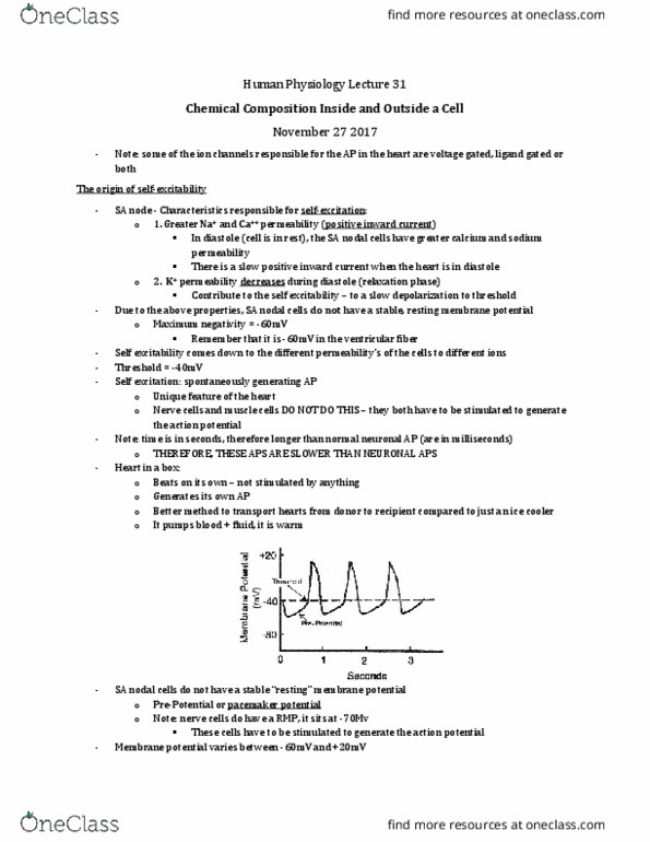
SA and AV Node Action Potential
Phase 0- Depolarisation
• Opening of voltage-gated Ca2+ channels causes depolarisation
Phase 3- Repolarisation
• Opening of voltage-gated K+ channels causes depolarisation
Phase 4- Spontaneous Depolarisation
• Funny currents occur due to hyperpolarisation-activated sodium channels in
the SA node
• This accounts for the SA node’s automaticity
Arrhythmia
• Abnormal site of impulse formation and/or pattern of conduction
• Arrhythmia classification:
− Site of origin of abnormality (atrial, junctional, ventricular)
− Heart rate- bradycardia or tachycardia
• Causes of arrhythmia:
− Ischaemia
− Fibrosis and damage to heart disrupts electrical balance
− Stress (increased SNS activity)
− Drugs
− Electrolyte imbalance
• Arrhythmias are common in patients who are:
− Treated with digoxin
− Under general anaesthesia
− Suffering from AMI
Class I Antiarrhythmic
• They are use-dependent Na+ channel blockers, and they selectively block:
find more resources at oneclass.com
find more resources at oneclass.com
Document Summary
Ion exchange changes electrical properties of cell, which increases/decreases cell excitability. Increasing excitability increases arrhythmia risk: when voltage-gated calcium channels open, ca2+ flows in, increasing contractility, excitability, and automaticity, sodium flows in- increases excitability, potassium flows out- decreases excitability. Sodium-potassium pump: transporter protein, uses atp to pump 2 k+ into the cell, and 3 na+ out of the cell. The long refractory period is protective from re-excitation during a heartbeat. Phase 0- rapid depolarisation: voltage-gated na+ channels open rapidly at threshold (~-70mv), causing na+ influx, this causes membrane depolarisation, the channels are open only for a few milliseconds. Phase 1- partial/early repolarisation: voltage-gated na+ channels close- inactivation of inward na+ current, additional k+ channels open briefly, the membrane transiently repolarises. Phase 2- plateau: l-type voltage-gated ca2+ channels open slowly, allowing the slow influx of ca2+ ions, there"s a small outward flow of k+ Phase 3- repolarisation: the ca2+ channels close, large increase in outward k+ current.