ANHB1102 Lecture Notes - Lecture 8: Anterior Interventricular Sulcus, Posterior Interventricular Sulcus, Superior Vena Cava

25 May 2018
School
Course
Professor

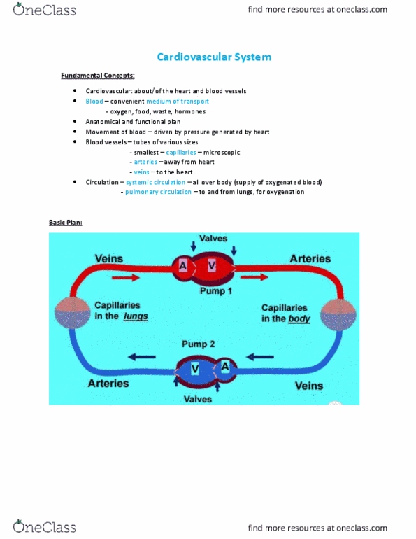
Cardiovascular System:
-The cardiovascular system consists of the heart and blood vessels. The heart is a muscular
pump that keeps blood flowing through the vessels which deliver oxygenated blood to all the
body’s organs then return deoxygenated blood to the heart.
Blood Vessels:
-Arteries: blood flow AWAY from the heart
-Veins: blood flow TOWARDS the heart
Circulation:
-Pulmonary circulation: to and from the lungs (for oxygenation)
-Supplied by the right atrium/ventricle
-Receives blood that has circulated through body
(deoxygenated)
-Pumps into pulmonary trunk which goes to the right and left
pulmonary arteries
-These transport blood to aveloli where gas exchange takes
place (becomes oxygenated)
-Flows through pulmonary veins to left side of the heart
-Systemic circulation: all over the body (supply oxygenated blood)
-Blood flows into the left atrium/ventricle
-Then flows through the aorta
-The aortic arch supplies oxygenated blood to the head, neck
and upper limbs
-The aorta travels through the thoracic and abdominal cavities to
supply other organs then supplies the lower limbs
-Deoxygenated blood then returns to the heart (right atrium/
ventricle) through the superior vena cava (draining everything in
upper body) and inferior vena cava (draining everything below
diaphragm)
Location of the Heart:
-Heart is located in the thoracic cavity in the mediastinum
between the lungs and deep to the sternum
-It is titled towards the left (2/3 lie to the left of the median
plane)
-Apex of heart is immediately above diaphragm
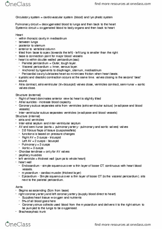
Pericardium:
-A double-walled sac that encloses the heart
-Tough, superficial fibrous layer of dense irregular
connective tissue
-Thin, deep serous layer which turns inwards at the base of the heart to form the visceral
pericardium
-Facilitates movement/expansion
-Anchored by ligaments to the diaphragm below and sternum anterior to it
-Parietal (outer) pericardium and Visceral (inner) pericardium
-Pericardial Cavity: formed between two layers and contains pericardial
fluid which is exuded by the serous layer of pericardiac sac
-Fluid lubricates membranes and prevents friction
Heart Wall:
-Consists of three layers:
-Epicardium (visceral pericardium): serous membrane of the external heart surface
-Mainly simple squamous epithelium overlying a thin layer of areolar tissue

-Can also include thick layer of adipose tissue
-Endocardium: lines interior of the heart chambers
-Simple squamous epithelium overruling thin areolar tissue
-No adipose tissue
-Covers valve surfaces and is continuous with endothelium of blood
vessels
-Myocardium: In between the Epicardium and Endocardium
-Cardiac muscle
-Thickest layer and performs the work of the heart
Gross Anatomy of the Heart:
-Chambers:
-Right atrium
-Left atrium
-Receive blood
-Right ventricle
-Left Ventricle
-Pump blood into arteries
and around body
-Boundaries of chambers
marked by sulci (grooves)
which are filled by fat and the
coronary blood vessels
-Coronary
(antrioventricular) sulcus:
separates atria from
ventricles
-Anterior interventricular
sulcus and Posterior
interventricular sulcus:
extend obliquely down the
heart from coronary sulcus
toward the apex

Valves:
-Atrioventriucular (AV) valves: regulates the openings between atria and ventricles
-Right AV valve: three cusps
-Left AV valve/Mitral valve: two cusps
-Chordae tendinae connect valve cusps to conical papillary muscles on the floor of the ventricle
-These prevent the AV valves from flipping inside out to bulging into the atria when the
ventricles contract
-Semilunar valves (pulmonary and aortic valves): regulate the flow of blood from the ventricles
into the great arteries
-Each has three cusps shaped like shirt pockets and do not have any chords tendinae
-Valves open and close by changes in blood pressure that occur as the heart chambers contract
and relax
-Pulmonary valve: controls opening from the right ventricle into pulmonary trunk
-Aortic valve: controls opening from left ventricle into the aorta
Coronary Vessels:
-Heart itself requires an abundant supply of oxygen, blood and nutrients
-The myocardium has its own supply of arteries and capillaries that deliver blood to every muscle
in the cell = coronary circulation
-Immediately after the aorta leaves the left ventricle, it fives off a right and left coronary artery
-Left coronary artery (LCA): travels through the coronary sulcus under the left auricle and
divides into two branches
1. Anterior interventricular branch: travels down the anterior inter ventricular sulcus to the
apex, rounds the bend and travels a short distance up the posterior side of the heart
where it joins the posterior interventricular branch/left anterior descending (LAD)
branch: supplies blood to both ventricles and the anterior two thirds of the inter
ventricular septum