ANHB2212 Lecture Notes - Lecture 3: Stratum Granulosum, Stratum Spinosum, Stratum Corneum

25 May 2018
School
Course
Professor

Embryology beyond the first 4 weeks:
Germ layers are the building blocks of organs:
Ectoderm – nerve and some epithelia
Mesoderm – all connective tissues, muscle and some
epithelia
Endoderm – some epithelia
Ectoderm:
Subdivisions:
- Ectodermal germ layer gives rise to the organs and structures that maintain contact
with the outside world.
- Ectoderm differentiates due to the formation of the neural groove and tube into two
main subdivisions.
Neuroectoderm: neural tissue precursor that forms the neural tube and crest.
Surface ectoderm: epidermis of skin, including hair, nails and glands.
Neural tissue is the first type committed to its fate at the end of week 3
- first sign is localized thickening of the ectoderm cranial to the primitive streak
- this is the neural plate.
find more resources at oneclass.com
find more resources at oneclass.com

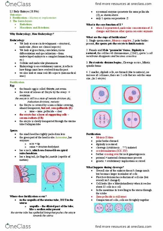
Surface Ectoderm:
Initially a single-cell layer thick –
proliferates and forms a new outer
layer – periderm (simple squamous
epithelium).
Under the periderm are proliferating
cells that form the basal layer (later to
be germative layer).
- Separated from the dermis by
the basement membrane.
By month 5 the periderm is shed and the intermediate layer is
replaced by 3 definitive layers of keratinocytes.
- Stratum spinosum – inner
- Stratum granulosum – middle
- Stratum corneum – outer
Epidermis also forms other specialized structures.
Surface specializations are derived from ectodermal layer and
formed in response to inductive signals from underlying
mesoderm.
- Hair follicles
- Nails
- Sebaceous and sweat glands
- Mammary glands
Other specialized structures.
- Lens of eye
- Lining of mouth
- Enamel of teeth
- Anterior pituitary
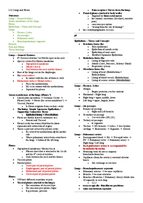
Neural tube ectoderm:
Gives rise to the CNS
- Brain is expanded cranial end of the embryo
- Neural canal forms ventricular system and
central canal of spinal cord.
- Nerve cell bodies line the central canal
- motor – ventral
- sensory – dorsal
find more resources at oneclass.com
find more resources at oneclass.com

Neural crest ectoderm:
Gives rise to peripheral nervous
system
Neural crest cells bud off and
migrate after being formed in
neurulation along pathways where
they differentiate.
- Dorsal root ganglia
- Autonomic ganglia
- Adrenal medulla
- Schwann cells
- Pia and arachnoid meninges
- Melanocytes
- Dentine and cornea
- Truncoconal septum of heart
Development of the head:
Many structures develop through interactions between the neural tube or crest and the surface
ectoderm.
- Cranial vault
- Ectodermal sensory placodes (ear, eye, nose)
- Pharyngeal arches
Some are a continuation of body structures
- Vertebral column
- Base of skull
Mesoderm:
Gives rise to all muscle, all connective tissue in body and limbs and most in the head, and
certain epithelia
Subdivided into distinct regions during end of week 3 into 4 as the body folds. Named according
to location on transverse axis of body.
Paraxial
By the end of week 3 is organized in to segments (somites).
- Appear in an ordered sequence cranial to caudal – 3 pairs per day until the end of the 5th
week (42-44 pairs total).
Somites differentiate into 3 components
find more resources at oneclass.com
find more resources at oneclass.com
Document Summary
Germ layers are the building blocks of organs: Mesoderm all connective tissues, muscle and some epithelia. Ectodermal germ layer gives rise to the organs and structures that maintain contact with the outside world. Ectoderm differentiates due to the formation of the neural groove and tube into two main subdivisions. Neuroectoderm: neural tissue precursor that forms the neural tube and crest. Surface ectoderm: epidermis of skin, including hair, nails and glands. Neural tissue is the first type committed to its fate at the end of week 3 first sign is localized thickening of the ectoderm cranial to the primitive streak this is the neural plate. Initially a single-cell layer thick proliferates and forms a new outer layer periderm (simple squamous epithelium). Under the periderm are proliferating cells that form the basal layer (later to be germative layer). Separated from the dermis by the basement membrane.