ANHB2212 Lecture Notes - Lecture 9: External Intercostal Muscles, Pulmonary Pleurae, Intercostal Muscle

25 May 2018
School
Course
Professor

Mechanisms of Breathing:
Breathing:
Quiet breathing: relaxed, unconscious, automatic
- uses diaphragm and intercostals
Forced breathing: unusually deep or rapid breathing
- diaphragm, intercostals and accessory muscles
lungs don’t ventilate themselves, only muscle contained within is smooth in the walls of the
bronchi and bronchioles
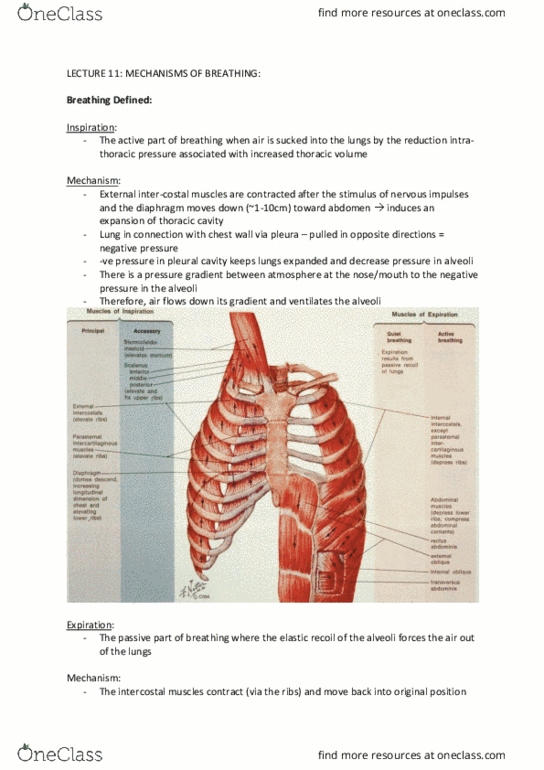
Inspiration – active part of breathing when air is sucked into the lungs by the reduction in intra-
thoracic pressure associated with increased thoracic volume.
Mechanism:
External inter-costal muscles contracted after stimulus of nervous impulses and the
diaphragm moves down (-1-10cm) toward the abdomen – induces expansion of the thoracic
cavity.
Lung is in connection with the chest wall via the pleura (parietal lining the rib cage, visceral
constituting lung surface) – via the pleura the lungs and chest wall are pulled in opposite
directions.
Results in negative pressure in the pleural cavity
keeping the lungs expanded and decrease pressure in
the alveoli – there is a pressure gradient between the
atmosphere at the nose/mouth to the negative pressure
in the alveoli.
Air thus flows down gradient and ventilates the alveoli.
find more resources at oneclass.com
find more resources at oneclass.com

Expiration: passive part of breathing where the elastic recoil of the alveoli forces the air out of
the lungs
Mechanism:
Intercostal muscles contract (via ribs) and move back into their original position
Diaphragm moves back, reducing space in thoracic cavity, and increasing pressure in lung
Recoil of thoracic cage compresses lungs and raises intra-pulmonary pressure (+1cm H20)
In forced breathing the accessory muscles raise the pressure substantially (+40cm H20).
Anatomy of chest wall:
Thoracic cage: skeleton of the chest that comprises the thoracic vertebrae, ribs and sternum.
- Ribs and sternum form rib cage that supports the walls of the thoracic cavity
- Is broad inferiorly, narrow superiorly and relatively flattened in the anterior-posterior
plane.
2 primary functions:
1) Protection for structures in thoracic cavity
2) Attachment for muscles involved with – respiration
- movement of vertebral column
- movement of pectoral girdle and upper limbs
Regional anatomy:
12 thoracic vertebrae
Sternum
12 ribs on each side and respective costal cartilage.
find more resources at oneclass.com
find more resources at oneclass.com

Thoracic inlet: superior opening (aperture) of thoracic cage.
- Approx. 5cm anteroposteriorly and 10cm transversely
- Bounded by superior border of the manubrium, posteriorly by the body of T1 and
laterally by the inner border of the 1st ribs.
Thoracic outlet: inferior opening (aperture)
- bounded posteriorly by T12, posterolaterally by 12th ribs, anteriorly by xiphoid process
and anterolaterally by costal margin.
Costal margin: portion of the inferior edge of the thorax defined by the articular cartilages of
ribs 7-10 (false ribs).
- Forms large inverted V-shape on the inferior border.
True ribs – 1-7, articulate directly via separate costochondral cartilages to the sternum.
- Gradually increase in length and radius of curvature
False ribs – 8-10, fuse together before reaching the sternum (shared costochondral cartilage)
Floating ribs – 11 and 12, don’t articulate with sternum,
Joints of ribs with sternum:
Via costal cartilage.
- Extends medially from anterior rib ends and articulate with the sternum (ribs 1-5) or
costal arch (ribs 6-10).
- Show the typical structure of hyaline cartilage – precursor of bone, widely dispersed type
II collagen fibres.
- Bound to sternal end of the rib by continuity of the periosteum of the bone and
perichondrium of the cartilage.
- Semi-moveable joint that permits flexibility in the rib cage whilst keeping the ribs
connected to the sternum
- Allows rib cage to expand with the lungs during deep inhalation
- Also serves to some degree as a shock absorber to anterior blows to the thoracic cage
- Remember about age related ossification of the cartilage.
First rib – synchondrosis – little flexibility, when rib elevated manubrium tilted upward
2-7 ribs – synovial sternocostal joints – allow slight gliding motion.
Manubrium and sternum – synchondrosis – as manubrium tilts the body of sternum doesn’t.
- Sternal angle increases with respiration
Body and xiphoid process – synchondrosis
find more resources at oneclass.com
find more resources at oneclass.com
Document Summary
Diaphragm, intercostals and accessory muscles lungs don"t ventilate themselves, only muscle contained within is smooth in the walls of the bronchi and bronchioles. Inspiration active part of breathing when air is sucked into the lungs by the reduction in intra- thoracic pressure associated with increased thoracic volume. External inter-costal muscles contracted after stimulus of nervous impulses and the diaphragm moves down (-1-10cm) toward the abdomen induces expansion of the thoracic cavity. Lung is in connection with the chest wall via the pleura (parietal lining the rib cage, visceral constituting lung surface) via the pleura the lungs and chest wall are pulled in opposite directions. Results in negative pressure in the pleural cavity keeping the lungs expanded and decrease pressure in the alveoli there is a pressure gradient between the atmosphere at the nose/mouth to the negative pressure in the alveoli. Air thus flows down gradient and ventilates the alveoli.