ANHB2212 Lecture Notes - Lecture 5: Spinal Nerve, Myotome, Somite

25 May 2018
School
Course
Professor

Development and Growth of the Vertebral Column:
Somites:
Notochord induces segmentation
Mesoderm condenses into blocks called somites
- Dermatome – skin of the back
- Myotome – segmented muscles that act on either side of the notochord to swim (in
primitive chordates).
- vertebral muscle
- Sclerotome – segmented bony parts
- vertebrae.
1 SOMITE DOESN’T MAKE 1 VERTEBRA
vertebral muscles are segmental
- 1 muscle = 1 myotome segment
But if muscles are to move the column, each must attach to 2 vertebrae.
Each vertebra forms two adjacent segments (sclerotomes).
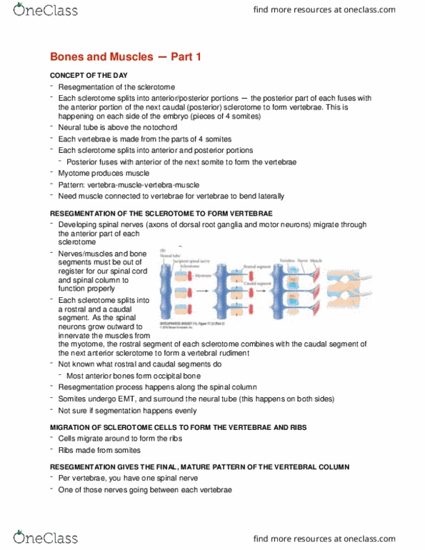
Re-segmentation:
Early on, sclerotomes divide into cranial and caudal halves.
- Caudal end of one segment fuses with cranial end of segment behind it.
Body wall vessels lie
between somites
- Intersegmental
arteries.
These provide better
nutrition to perinotochordal
cells nearby.
- Perinotochordal cells
near arteries grow
larger and become vertebral bodies.
- Less nourished parts in between become discs.
find more resources at oneclass.com
find more resources at oneclass.com

- Notochord is squeezed into the zone
between the developing bodies.
Vertebral development
3 stage process:
Mesenchyme →cartilage→bone
- Endochondral ossification
Mesenchymatous stage – week 4-6
Sclerotome cells migrate
- Perinotochordal sheath (centrum)
- Neural arch
- Costal element
-
Cartilaginous stage – week 6-9
Begins with mesenchymatous scaffold of the
vertebra
Paired chrondrofication centres appear:
- Centrum
- Neural arch
- Costal
Mesenchyme gradually replaced by hyaline
cartilage
find more resources at oneclass.com
find more resources at oneclass.com

In region where discs form, fibrocartilage forms a ring
(annulus fibrosus) around the notochordal element
(nucleus pulposus).
- Discs in line with rest of somite.
Anomalies in cartilaginous stage:
Because cartilage centres are paired, if one side fails
to form, vertebrae develop asymmetrically.
- Hemivertebrae
- “failure of formation”
Osseous stage: week 8 onwards
Begins with cartilage model of vertebrae
Centres of ossification appear
- Centrum (unpaired)
- Neural arches
- Costal elements – either fuse with
rest of vertebra, or become ribs and
develop joints
(thorax).
Bone grows, but cartilage growth plates
continue to separate the ossification centres
- Neurolaminar
find more resources at oneclass.com
find more resources at oneclass.com
Document Summary
Myotome segmented muscles that act on either side of the notochord to swim (in primitive chordates). 1 somite doesn"t make 1 vertebra vertebral muscles are segmental. But if muscles are to move the column, each must attach to 2 vertebrae. Early on, sclerotomes divide into cranial and caudal halves. Caudal end of one segment fuses with cranial end of segment behind it. These provide better nutrition to perinotochordal cells nearby. Perinotochordal cells near arteries grow larger and become vertebral bodies. Less nourished parts in between become discs. Notochord is squeezed into the zone between the developing bodies. In region where discs form, fibrocartilage forms a ring (annulus fibrosus) around the notochordal element (nucleus pulposus). Discs in line with rest of somite. Because cartilage centres are paired, if one side fails to form, vertebrae develop asymmetrically. Costal elements either fuse with rest of vertebra, or become ribs and develop joints (thorax).