ANHB2212 Lecture Notes - Lecture 17: J. P. Dutta, Falciform Ligament, Aorta

28 May 2018
School
Course
Professor

L17 Embryology – Digestive System
Outcomes
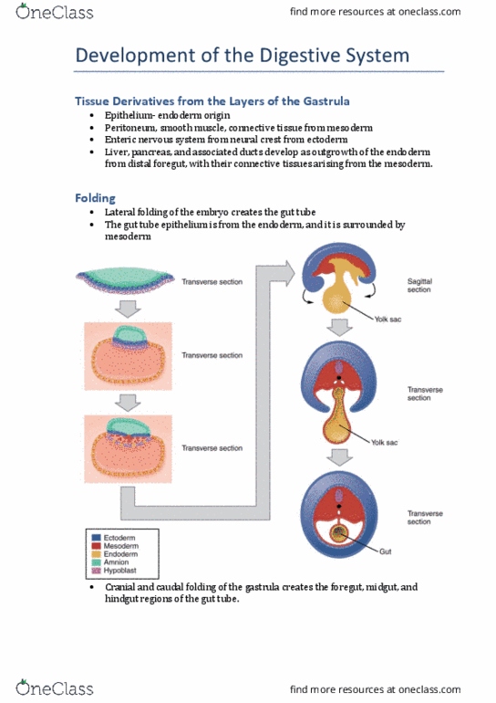
Head and tail folds
• Cranial end of gut tube
• Caudal end of gut tube
Lateral folds
• Coelomic cavity → peritoneal cavity → etc
o Is lined by peritoneal epithelium
▪ Mesodermal in origin
o Mesentery at dorsal border
▪ 2 Epithelial layers, connective
tissue in between
• Endoderm gives rise to lining epithelium of
digestive tube and secretory tissues of digestive
systems (Lamina Propria, submucosa etc)
• CT + Muscles = always mesodermal in origin
yolk sac gets smaller and smaller
Mesenteries **
• FMH
• Foregut has 2 mesenteries
o VM + DM
• Vitello – intestinal duct is located at the midpoint
of midgut
• Ventral mesogastrium connects foregut to anterior
abdominal wall
• Dorsal mesogastrium connects foregut to posterior
abdominal wall
• Dorsal mesogastrium is continuous in FMH
• Umbilical vein joins ventral mesogastrium to bring
deoxygenated blood to the heart
Foregut gives rise to
1. Abdominal oesophagus
2. Stomach
3. Cranial half of duodenum
Foregut mesenteries
• VM and DM, right? That’s fine
• Liver develops in Ventral mesogastrium
o Has 2 parts
o 1 part (Falciform ligament to anterior
abdominal wall)
o 1 part (Lesser omentum to lesser
curvature of stomach)
• Spleen develops in dorsal mesogastrium
o 3 parts
o gastrosplenic ligament
o gastrophrenic ligament
o linorenal ligament
Midgut
• superior mesenteric artery (from aorta) extends to
midgut, to yolk sac and runs towards the apex of
the midgut loop
• yolksac is now small and vitello-intestinal duct is at
the tip of midgut loop
• Cranial limb / Pre-arterial
• Caudal limb / Post-arterial
o Caecal bud
Midgut gives rise to
1. Caudal duodenum
2. Jejunum
3. Ileum
4. Caecum
5. Appendix
6. Ascending colon
7. 2/3 transverse colon
Midgut – Rotation of the limbs
• Cranial <-> Caudal
• X = junction of foregut/midgut
• Cranial limb gives rise to small intestines
• Caudal limb gives rise to ascending colon
• Don’t forget about the V-I duct at the apex of the
midgut
• When the midgut loop returns to the abdomen, the
structures are initially in a subhepatic location
o Caecum = subhepatic
o VI duct = close to caecum
Hindgut gives rise to
1. Splenic flexure
2. Descending colon
3. Sigmoid colon
4. Rectum
5. Part of anal canal
Anal canal
• Opens from clocal membrane
• The hindgut gives rise to the epithelial part of the
upper part of the anal canal
• Ectodermal depression (proctodeum) is the lower
part of the membrane
o That disappears as hindgut epithelial
(endodermal lining) joints ectodermal/
proctodeum
find more resources at oneclass.com
find more resources at oneclass.com