HUMB1001 Lecture Notes - Lecture 6: Loose Connective Tissue, Uric Acid, Peritubular Capillaries

2 Aug 2018
School
Department
Course
Professor
Document Summary
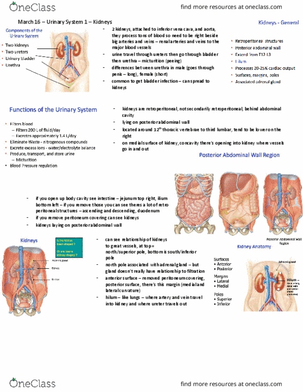
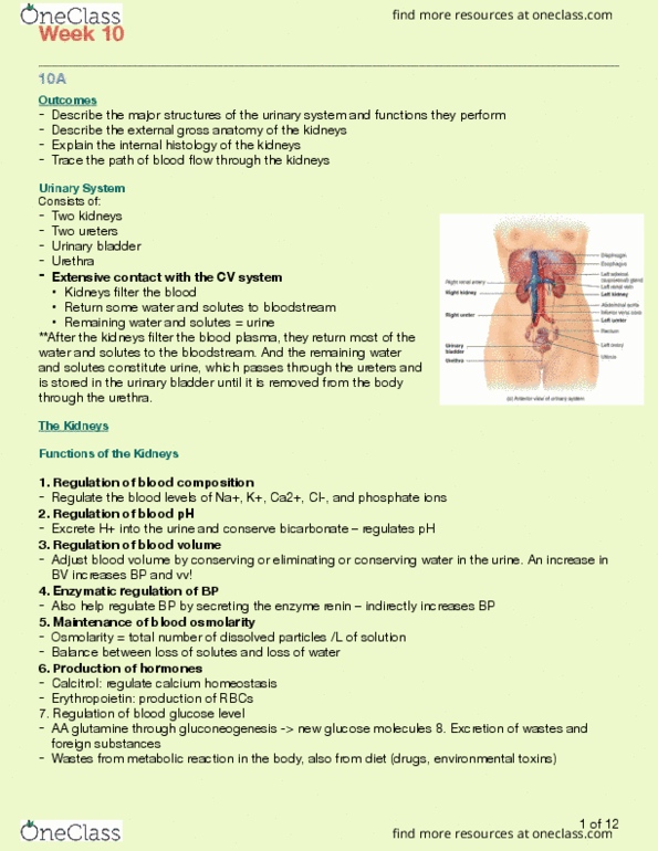
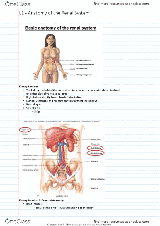
The kidneys are two bean-shaped organs (the size of the fist, ~ 130g). Renal capsule: fibrous connective tissue surrounding each kidney. Adipose tissue: engulfs renal capsule and acts as cushioning. Renal fascia: thin layer loose connective tissue, which anchors kidneys to posterior abdominal wall. The renal hilum carries blood vessels and nerves into the kidney and serves as the point of attachment for the ureters: the renal artery and nerves enter, the renal vein and ureter exit. The hilum opens into the renal sinus: a cavity filled with fat and connective tissue. Renal cortex: outer area in the kidney which contains the renal corpuscle and the proximal and distal convoluted tubules of the nephron. Renal columns: sections of cortical tissue that extend down into the medulla separating the renal pyramids. Medulla: inner area in the kidney which surrounds the renal sinus and contains the loop of henle and the collecting duct of the nephron.