BIOL10003 Lecture 4: Medical Mycology

21 Jul 2018
School
Department
Course
Professor

Textbook: Chapter 30
Hence very difficult to treat
Fairly basic in terms of appearance. Earlier branches (Chytridiomycota) = mixture of different
organisms. No human pathogenic known today for chytrid. Whereas the other 3 cause diseases.
Mycoses
1.
Toxic fungi
2.
Allergens
3.
Drugs from fungi
4.
FUNGI IN MEDICINE:
Human Mycoses
1.
Fungal disease in humans is common (usually not life-threatening)
-
Immune system is effective
○
Few species cause few diseases in mammals. Need to overcome below factors to cause
disease
-
Medical Mycology
Tuesday, 1 August 2017
9:43 PM
Week 2 Page 1

○
Grow slowly under low oxygen
○
Few are capable of growth at 37◦C
○
There are many fungal diseases of plants
-
Common element is growth at 37◦C
○
Virulence (degree of damage caused by a microbe to its host) "factors" vary between species
-
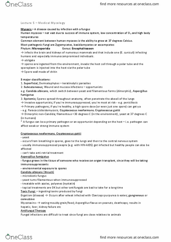
e.g. of human mycoses = Microsporidia (phylum Microspora)
700 species
-
Encephalitozoon spp. Important parasite of humans
-
Colonies multiple organs, including brain, kidney, liver, adrenal glands, optic nerves,
myocardium (muscular tissue of heart)
-
Minute, obligate (parasitic organism that cannot complete its life-cycle without exploiting a
suitable host. If no host, will fail to reproduce) intracellular parasites
-
Lack normal mitochondria and no flagella
-
Originally thought to be very primitive eukaryotes, now known to be a type of fungi
-
Spores ingested from environment
-
Invade host cell by everting structure known as polar tube
-
Sporoplasm (parasite cell without wall) is injected through the tube into the host cell
-
Spore wall is made of chitin (same as fungal wall) and stains with Calcofluor
-
MICROSPORIDIA (PHYLUM MICROSPOA):
Spore shot out filament that infects the cell.
3rd pic=inside part is spat out and injects itself into host. Cell wall is chitin. Polar tube is
wrapped around (little circles)
Encephalitozoon cuniculi was the second fungus sequenced (1999, although no one
realised it was a fungus)
○
Enterocytozoon bieneusi is a common cause of diarrhea amongst children in African
countries with high AIDS incidence
○
Microsporidiann life cycle:
-
i.e. which species causes which disease
Can be classified based on taxonomy
-
HUMAN MYCOSES:
Week 2 Page 2

i.e. which species causes which disease
○
Superficial - on the surface of skin or nail
a.
Subcutaneous - below surface
b.
Some species can cause disease in more than one location
i.
Systemic infections - growing throughout body
c.
Three major classifications based on the location of the disease
-
Unlikely to cause death
-
Genomes are specialised to life on animals - have keratinase to break down keratin, can
break down lipid
-
Parasites (not saprophytes)
-
Keratinolytic
-
Superficial infections - Dermatophytes
a.
e.g. Ringworm, tinea
etc.
Trichophyton spp.,
Microsporium spp.,
Epidermophyton spp.
Pityriasis versicolor
(common skin
disease caused by an
overgrowth of the
yeast fungus called
Pityrosporum
orbiculare
(Malassezia furfur).
Most adults have
Pityorospporum
orbiculare on their
skin: however, in a
few people its
presence results in a
harmless skin disease
Malassezia furfur - need human lipid to grow on our skin.
Tinea pedis or
athlete's foot -
caused by
Epidermophyton
floccosum
Asexual pores being produce
Onychomycosis or
nail fungus
Fungal infection that causes fingernails or toenails to thicken,
discolour, disfigure, and split.
-
These fungi are not really parasitic in the sense of attacking living tissue. They attack the dead
cells of the epidermis, and cause a kind of dermatitis. The irritation caused by the fungus
stimulates the skin cells to divide more rapidly. This means that more flakes of skin containing
infective mycelium will be shed.
Subcutaneous - wound and mucosa infections opportunists
b.
Enter via wound
-
Lesions ulcerated, crusted
-
Can spread through lymph system
-
Subcutaneous mycoses
Phialophora,
Cladosporium,
Sporothrix,
Acremonium
Thrush
Candida albicans,
Week 2 Page 3
Document Summary
Earlier branches (chytridiomycota) = mixture of different organisms. Fungal disease in humans is common (usually not life-threatening) Need to overcome below factors to cause disease. Virulence (degree of damage caused by a microbe to its host) factors vary between species e. g. of human mycoses = microsporidia (phylum microspora) Colonies multiple organs, including brain, kidney, liver, adrenal glands, optic nerves, myocardium (muscular tissue of heart) Minute, obligate (parasitic organism that cannot complete its life-cycle without exploiting a suitable host. If no host, will fail to reproduce) intracellular parasites. Originally thought to be very primitive eukaryotes, now known to be a type of fungi. Invade host cell by everting structure known as polar tube. Sporoplasm (parasite cell without wall) is injected through the tube into the host cell. Spore wall is made of chitin (same as fungal wall) and stains with calcofluor. Spore shot out filament that infects the cell. 3rd pic=inside part is spat out and injects itself into host.