NEUR30003 Lecture Notes - Lecture 7: Lateral Geniculate Nucleus, Low Frequency, Helicotrema

10 May 2018
School
Department
Course
Professor

Lecture 7
•
•
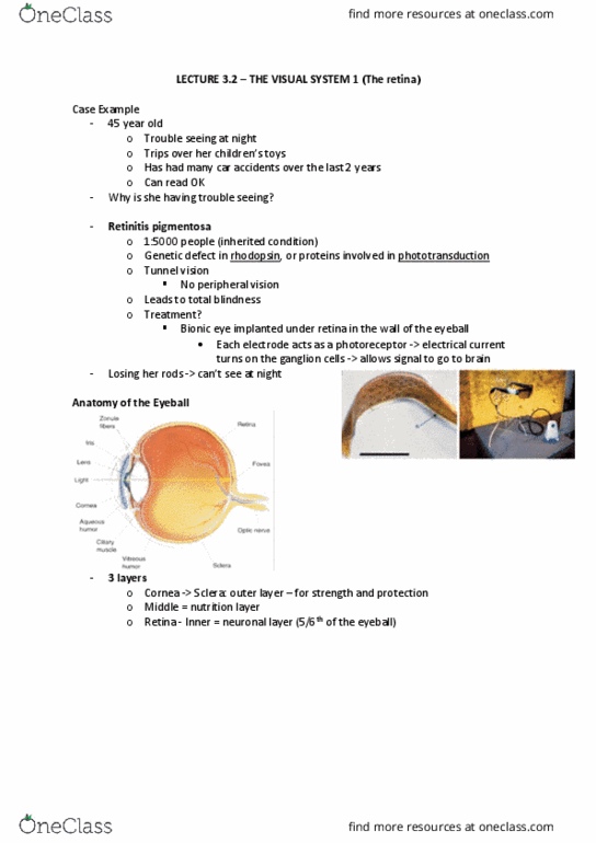
• Light goes in from the front through the cornea → bent by lens → focuses on back
part of eye (retina, the most internal surface of eyeball, nervous layer [layer of
neurons that allow us to pick up light etc])
• Optic nerve collects information from retina and passes neural info down to brain
• Axons of retinal ganglion cells within optic nerve
• Outside layer [white of eye, sclera] → tough, strong
• Middle layer (vasculature)
• Inside layer (retina, allows us to see)
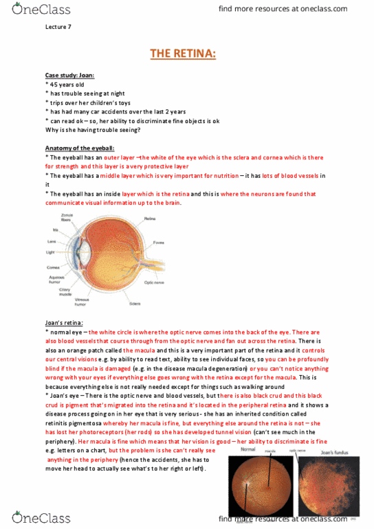
• Macula: darker part of retina, centre of macula is fovea [area that allows us to see
centrally, critical for vision, for good high resolution vision, ability to see fine detail]
find more resources at oneclass.com
find more resources at oneclass.com

•
• Retinitis pigmentosa: inherited condition which causes gradual loss of broad
photoreceptors in eye, lose peripheral vision until tunnel vision and gets smaller and
smaller
• Neural factors and optical factors limit visual acuity (ability to see fine detail)
• Optical factors affecting visual acuity: pupil size [changes based on light level,
smaller → see more clearly], clarity of optical media (cataracts [opaque lens], corneal
opacities [scars etc]), refractive errors [how long the eyeball is] → blur (myopia
[short-sightedness, eyeball is too long, light doesn’t focus on the correct place behind
eyeball, focus too early → what happens at retina is blurry], hypermetropia [long-
sightedness, eyeball is too small], astigmatism [eyeball is the wrong shape],
presbyopia [issue with lens])
• Refractive error is important for creating blur and having factor in how well one can
see fine detail
find more resources at oneclass.com
find more resources at oneclass.com

•
• Light passes through all the layers of retina until hits photoreceptors which pick up
light and change it to neural signal
•
• Rods and cones - named after shape
• Rods: many, allow us to see at night, very sensitive to light, main photoreceptor
(95%), occupy most outside part of retina [outside of eye, deep in wall of eyeball, sit
next to outer layers of eyeball]
find more resources at oneclass.com
find more resources at oneclass.com
Document Summary
Info from nasal retina (communicated by ganglion cells there) nasal fibres cross at the chiasm synapse in the opposite lgns of each side of brain. If lesion on same side in both eyes defect must arise from post chiasm. Sound waves of varying pressure propagate through the air to reach the ear, reaching the outer ear propagates down the auditory canal producing vibrations in the tympanic membrane. Three middle ear bones or ossicles, the malleus, incus and stapes, transfer vibrations efficiently from the tympanic membrane (air) to the oval window (liquid) by amplifying pressure. The eustation tube allows equalisation of pressure across the tympanic membrane: 2. Within the inner ear the coiled cochlea contains two liquid filled chambers, the scala vestibuli and scala tympani (low potassiumion concentration), separated by the cochlear duct (scala media high potassium ion concentration) containing the basilar membrane. The organ of corti sits on top of the basilar membrane with many mechanically supporting cells.