NEUR30003 Lecture Notes - Lecture 4: Viscoelasticity, Neurological Examination, Gyrus

10 May 2018
School
Department
Course
Professor

Lecture 4
1. Before high resolution methods of investigating neural strucutre and function, the
correlations between these proporties was evident in the clinicial examination of neural
disorders and the subsequent gross morphology of the NS. Examination of the spinal and
cranial nerve function in a clinical setting is still a fundamental part of neurological
examination.
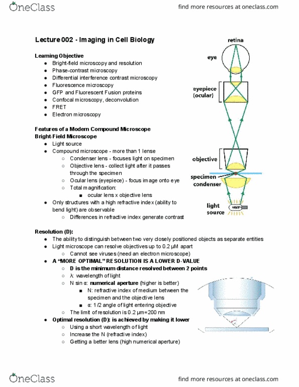
2. The microscopic study of brain tissue has revealed the variety of neuronal forms. Imaging
by microscopes (or telescopes, or eyes for that matter) is fundamentally limited by the
wavelength of the light. The wavelength of electrons allows resolution down to atomic
dimensions, but visible light wavelengths limited the maximum achievable resolution to about
250 nm. The location of objects smaller than this can be achieved if the objects give off light
(as in fluorescence microscopy) but even then, the actual location of these light sources is
obscured due to diffraction by the optics. The key to any imaging or measuring technology's
application is resolution - the spatial resolution of conventional light microscopes is governed
ultimately by the wavelength of visible light, the spatial resolution of electron microscope is
determined by the wavelength of the electron beam.
3. Very recently, several microscopy techniques have successfully overcome this diffraction
limit. These new methods depend on controlling the activation and deactivation of
fluorescent molecules. In stimulated emission depletion (STED) methods, super resolution is
achieved by narrowing the point spread function the diffraction disc (Airy disc) by using a
laser to precisely deactivate the outmost portion of the disc. In stochastic optical
reconstruction (STORM) the random switching of fluorescence of individual molecules allows
their individual contributions to be imaged: single molecules, which are stochastically
switched on, imaged and localized, and then switched off. Many cycles of this allows the
centre of these discs to be calculated and the image is constructed from millions of locations.
4. The discovery of the nerve impulse and the subsequent characterization of its electrical
properties lead to an understanding of how neurons produce and propagate and integrate
electrical signals. Microelectrodes are used to record the activity of single neurons; these have very
good spatial (i.e. single cell, and even single membrane channel) resolution and also very fast
temporal resolution (sub-microsecond). Combining molecular biology with high-resolution
electrophysiology, such as patch - clamp electrodes, can explicitly define the molecular basis
of electrical activity of neurons.
5. The scope or scale of these techniques is also important; microscopes have great spatial
resolution but can only be used on single or small groups of neurons or small regions of tissue - other
techniques exist to allow whole regions or even whole brains to be visualised. Computer assisted x-
ray tomography (CT scans) and magnetic resonance imaging (MRI) can image structure in the brain
of living people, and functional MRI, positron emission computed tomography (PET) and
magnetoencephalography (MEG) scans can localise areas of increased brain function associated with
performance of tasksWhole brain imaging technologies utalise different electromagnetic
radiation. These techniques differ in several key parameters: spatial resolution; temporal
resolution (for functional measurements) and invasiveness (how much damage they might
do to the subject being imaged).
Notes on Brain Imaging technology
• CT or CAT scans (computed /computer assisted x-ray tomography) A beam of x-rays
passes through the brain. As it does so is attenuated slightly because it has hit dense
living tissues on the way through. Attenuation of the x-ray comes from the density of
the tissue encountered along the way. Dense tissue like bone greatly attenuates x-
rays; the grey matter of the brain causes less attenuation, and fluid even less. X-ray
detectors positioned around the circumference of the scanner collect attenuation
readings from multiple angles. A computer reconstructs an image of each slice. X-
rays are damaging to tissue, but CAT scans use relatively small doses. A typical
find more resources at oneclass.com
find more resources at oneclass.com

scan gives the same exposure as 8 months normal background radiation present in
the environment.
• PET (positron emission computed tomography) When radio-labelled compounds are
injected in trace amounts, their photon emissions can be detected much like x-rays in
CT. The images made represent the accumulation of the labelled compound. The
compound may reflect, for example, blood flow, oxygen or glucose metabolism, or
chemical concentrations (such as administered drugs), thus can provide functional or
pharmacological data. The ionising radiation used in PET and SPECT studies is
harmful in large doses; this limits the number of scans an individual can receive.
• MRI When protons are placed in a magnetic field, they oscillate - they wobble. The
frequency at which they oscillate depends on the strength of the magnetic field.
Protons are capable of absorbing energy if exposed to electromagnetic energy at the
frequency of oscillation. After they absorb energy, the nuclei release or reradiate this
energy so that they return to their initial state of equilibrium. This re-radiation or
transmission of radio frequency energy by the nuclei over time as they return to their
initial state is what is observed as the MRI signal. The great majority of protons in
tissues are those of water molecules, and thus the concentration of water in a region
produces a significant signal. The magnetic fields used are very strong -30,000-
60,000 times the earth's magnetic field strength but not at all harmful to tissues.
• fMRI Functional magnetic resonance imaging (fMRI) can determine the
neurobiological correlate of behaviour by identifying the brain regions that become
"active" during the performance of specific tasks. When a region of the brain
increases its activity there is an increase in the blood supply to the brain locally,
ensuring an adequate supply of oxygen to more active regions. In fact, the increase
in blood supply to active regions more than meets the increased oxygen demands,
as a result, the blood in active regions has higher oxygenated to de-oxygenated
blood. The properties of the protons of water molecules in the brain change slightly
between areas that are near blood with its oxygen exhausted relative to those near
freshly oxygenated blood. The difference in the MRI signal is very small (about2%)
and typically many repetitive trials of strictly repeated tasks are required to generate
clear patterns. The "blood oxygenation level dependent" (BOLD) imaging technique
does not measure tissue perfusion or flow directly but exploits the difference in the
signal decay of de-oxygenated blood.
• MEG Magnetoencephalography, or MEG, is a method of measuring the tiny magnetic
fields produced when groups of the brain's 100 billion or so cells, or neurons, are
active. These magnetic fields generated by electrical currents produced by coherent
neural activity are a billion times smaller than Earth's magnetic field (and 10,000
times smaller than the field surrounding a household wire). The most sensitive
magnetic detectors (semi quantum interference devices - SQUIDS) need to be
cooled to the temperature of liquid helium to detect these tiny fields. As MEG
provides no structural information, the MEG data are combined with MRI or CT
maps.
• How should the brain be studied?
• Neuroscientists are usually seeking reductionist, causal, mechanistic explanations of
how system components interact to produce system behaviour
• A key part of neurological diagnosis is to localise the location of damage by
examination of site-specific functions
find more resources at oneclass.com
find more resources at oneclass.com

•
• Can examine structure of nervous system - how to reveal dysfunction
• Segmental reflexes: tap on tendon → knee-jerk response
• Primitive reflexes: responses we shouldn’t see, present in babies, get supplanted as
we develop (something goes wrong in nervous system → primitive reflexes take
charge)
• Pin prick - noxious stimulus
• Cotton ball - light touch
•
find more resources at oneclass.com
find more resources at oneclass.com
Document Summary
Lecture 4: before high resolution methods of investigating neural strucutre and function, the correlations between these proporties was evident in the clinicial examination of neural disorders and the subsequent gross morphology of the ns. Examination of the spinal and cranial nerve function in a clinical setting is still a fundamental part of neurological examination: the microscopic study of brain tissue has revealed the variety of neuronal forms. Imaging by microscopes (or telescopes, or eyes for that matter) is fundamentally limited by the wavelength of the light. The wavelength of electrons allows resolution down to atomic dimensions, but visible light wavelengths limited the maximum achievable resolution to about. The location of objects smaller than this can be achieved if the objects give off light (as in fluorescence microscopy) but even then, the actual location of these light sources is obscured due to diffraction by the optics. These new methods depend on controlling the activation and deactivation of fluorescent molecules.