PSYC10003 Lecture Notes - Lecture 16: Visual Cortex, Extrastriate Cortex, David H. Hubel

13 Jun 2018
School
Department
Course
Professor

11th Apr ‘18
MBB1 Week 6; Lecture 16 Notes
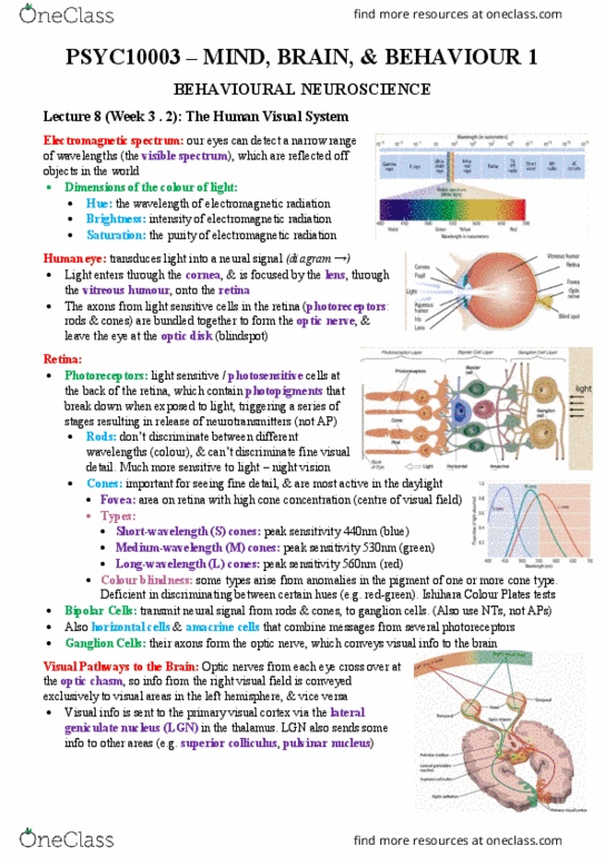
Receptive fields and the early visual pathway
-Hubel & Wiesel = mapped receptive fields in visual cortex
-measures the output of a singular isolated neuron
-placed electrode in Lateral Geniculate Axon of a cat
-presented the system with light stimuli
-when output is “perfectly the same” but it changed position, the system cannot convey that
change
-so orientation of visual stimuli doesn’t matter
-simple cell: cortical neuron that is linear in projection
-maps out visual field by drawing out excitatory and inhibitive neurons to see the difference
in orientation
-how we might generate orientation selectivity from in-selectivity
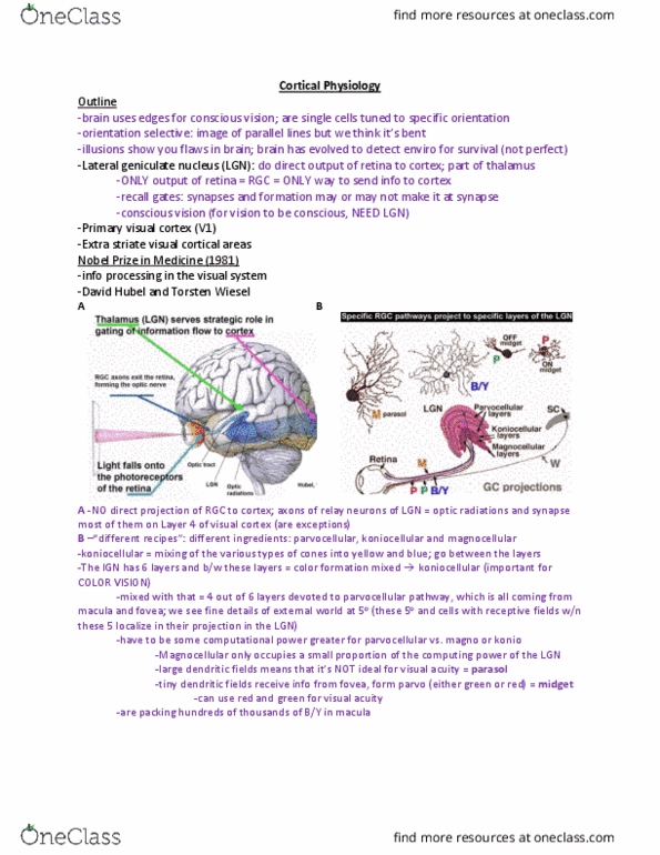
Dorsal Lateral Geniculate Nucleus
-retinal P cells: parvocellular
-retinal M cells: magnocellular
-koniocellular - high input from S cones
LGN
-“relay station”
-station post between primary visual cortex and retina
-more connections coming from cortex back to LGN
rather than LGN to cortex
-goes to 2 layers of LGN and then spreads into cortex
-input to cortical V1
-input to levels 4Cα(Magno) and 4Cβ (Parvo)
-right & left eyes input to adjacent cortical columns
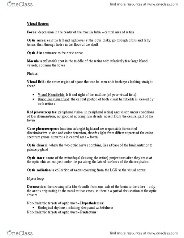
Primary visual cortex
-represents aspect of image but sparsely
-receptive fields selective for rudimentary properties of colour, motion, depth and form
-but some of these properties come via other input as well
-striate cortical arrangement
-geniculate input to layer IVc to PVC
-signals spread through other layers and then to extra-striate cortex
-if signal moves away from layer IV, receptive fields become more specialised in sensitivity
-more processing “machinery”
find more resources at oneclass.com
find more resources at oneclass.com
Document Summary
Hubel & wiesel = mapped receptive fields in visual cortex. Measures the output of a singular isolated neuron. Placed electrode in lateral geniculate axon of a cat. When output is perfectly the same but it changed position, the system cannot convey that change. So orientation of visual stimuli doesn"t matter. Simple cell: cortical neuron that is linear in projection. Maps out visual field by drawing out excitatory and inhibitive neurons to see the difference in orientation. How we might generate orientation selectivity from in-selectivity. Koniocellular - high input from s cones. Station post between primary visual cortex and retina. More connections coming from cortex back to lgn rather than lgn to cortex. Goes to 2 layers of lgn and then spreads into cortex. Input to levels 4c (magno) and 4c (parvo) Right & left eyes input to adjacent cortical columns. Receptive fields selective for rudimentary properties of colour, motion, depth and form.