PSY3710 Lecture Notes - Lecture 9: Halobacterium Salinarum, Electron Crystallography, Transmission Electron Microscopy

11 Apr 2018
School
Department
Course
Professor
Document Summary
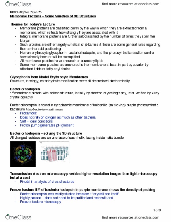
Class 9- membrane proteins some varieties of 3d structures. Membrane proteins are classified partly by the way in which they are extracted from a membrane, which reflects how strongly they are associated with it. Integral membrane proteins are further sub- classified by the number of ties they span the bilayer. Such proteins are either largely alpha- helical or beta barrel and there are some general rules regarding amino acid positioning. Human erythrocyte glycophorin, bacteriorhodopsin, and the photosynthetic reaction center have already been or will be exemplified. All membrane proteins have annular or boundary lipids. Some membrane proteins are anchored to the membrane at least in part by covalently- attached lipids to fatty- acrylic chains. Glycophorin from model erythrocyte membranes (structure, topology, carbohydrate modification were all determined biochemically) Bacteriorhodopsin (1st membrane protein with solved structure, initially by electron crystallography, later verified by x- ray crystallography. Bacteriorhodopsin is found in cytoplasmic membrane of halophilic (salt- loving) purple photosynthetic bacterium halobacterium salinarum.