BIOL 2005 Lecture Notes - Aorta, Heart Block, Small Cardiac Vein

30 Jan 2013
School
Department
Course
Professor
Document Summary
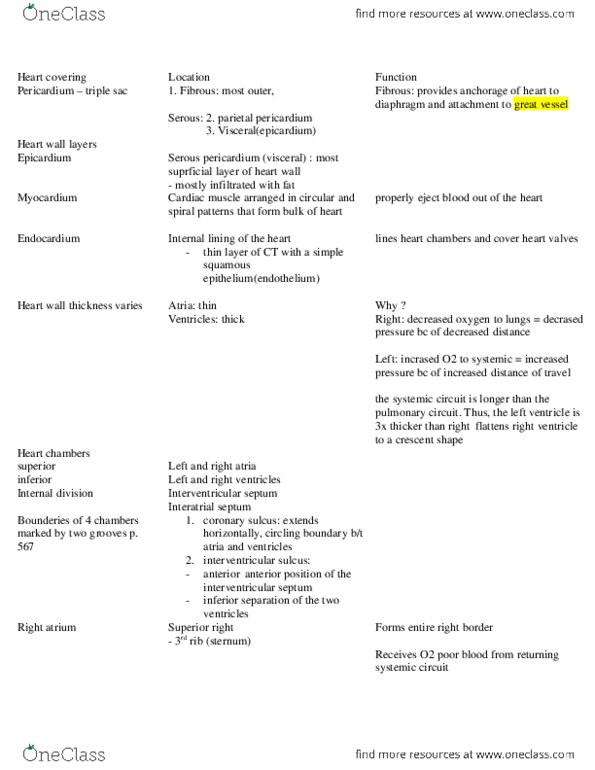
Heart: structure of outer layer (coelomic spaces) Coelomic sacs are cavities, lined by parietal and visceral serosa, and normally containing only serous fluid. Mesentery is the apposition of two serosal membranes, supporting an organ or gut. Pericardium is the coelomic sac around the heart. Inlet vessels (3): inferior vena cava, superior vena cava, coronary sinus. Musculi pectinate (aka pectinate muscles) ( primitive atrium) Tricuspid valve ( right ventricle: right ventricle. Septaomarginal trabeculae (aka moderator band) ( anterior papillary muscle) Anterior papillary anterior and posterior cusps of tricuspid. Posterior papillary posterior and septal cusps of tricuspid. Septal papillary anterior and septal cusps of tricuspid. Pulmonary valve ( pulmonary trunk lungs pulmonary veins left atrium: left atrium. Bicuspid valve (aka mitral valve) ( left ventricle) Nodules: be able to trace blood flow through the heart. Other important features of the heart: coronary vessels (understand which parts of heart each vessel go to) Small cardiac vein ( coronary sulcus: cardiac cycle.