MECH 430 Lecture Notes - Lecture 4: Ectodomain, Thyrotropin Receptor, Signal Peptide

9 Jun 2018
School
Department
Course
Professor

Lecture 4
➢ The Molecular Basis of Hormone Action
o Extracellular signals are recognized by specific recognized by
specific cell surface receptors
▪ Lock & key
o The activated receptors start a cascade of protein activation
o The target proteins responsible for altered cell properties are
turned on or off
▪ i.e. receptor activation triggers a feedback circuit that
shuts off the receptor or removes it from the cell surface
o cells receive multiple signals that are integrated to determine
overall response of the cell
o Signals are received by receptors and passed on through a chain
of signally proteins
▪ we need specific receptors to get specific messages sent into the nucleus, so that there is
no cross-talk
o Each cell receives a multitude of signals
o Often the signal is exponentially amplified during transmission and results in an on or off stage
o One or more of the signaling proteins interact with target proteins
▪ i.e. key regulatory proteins that determine the properties of a cell
o the modified target proteins alter the properties of the cell
➢ Based on the signal receptor pathways can be divided into 2 classes
o A: Pathways with cell-surface receptors
▪ On the cell membrane to receive a hormonal signal from the
aqueous medium → hydrophilic type molecules
o B: Pathways with intracellular receptors
▪ Associated with hydrophobic molecules, such as steroids and
prostaglandin → lipid-based hormones go into the plasma
membrane → they often need a carrier protein to keep them
stable in the blood and to diffuse into the cell where there is an
intracellular receptor to receive the signal
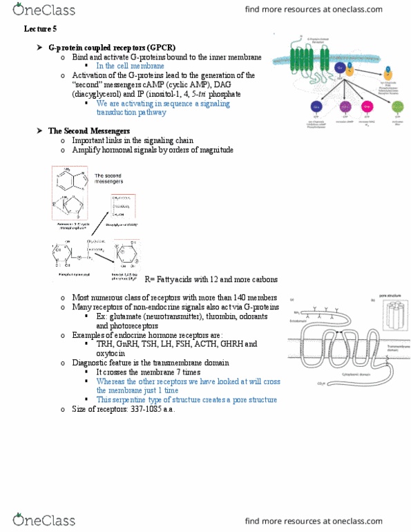
➢ Basic structure of a cell surface receptor
o Signal binds to ectodomain → NH2 end of peptide
▪ Has a specific shape to recognize in a lock and key manner
o Rich in cysteine residues → S-S bonds for folding & often glycosylated
o Hydrophobic transmembrane domain
▪ Alpha-helix
▪ May be more complex than shown
▪ Lipid soluble in the plasma membrane
o Cytoplasmic domain → CO2 end of peptide
▪ i.e. the endodomain → when this changes shape, it results in the changing shape of the
hydrophobic transmembrane domain
o The three domains are functionally independent → they are interchangeable
find more resources at oneclass.com
find more resources at oneclass.com

➢ Ectodomain may serve as a hormone binding protein
o Free ectodomain may circulate as a hormone binding
protein i
▪ The GH receptor ectodomain acts as a GH
binding protein in the blood circulation
• You can into equilibrium in free
circulation between GH bound and
aqueous phase of the blood circulation
▪ The ectodomain cleaved from the TSH
receptor may induce antibodies, which bind to
the receptor and mimic TSH action (cause of
hyperthyroidism in Grave’s disease
• Thus, the ectodomain can cause
problems
➢ The cytoplasmic domain of an activated receptor relays the signal to the interior of the cell
o The activated cytoplasmic domain induces a signaling cascade
▪ Relay of conformational changes of signaling proteins
o So the proteins keep changing shape according to their positions and state
o Such conformational changes are induced by
▪ Phosphorylation of proteins
▪ Binding between proteins
➢ Many signaling proteins are activated by phosphorylation at the amino acids serine, threonine and
tyrosine
• Hydroxyl group is phosphorylated in all 3 cases
• Tyrosine has a giant carbon projecting out of the backbone
o This docking site is where other hormones would interact with it at the surface of the
protein
find more resources at oneclass.com
find more resources at oneclass.com

➢ Protein phosphorylation
o The 3 a.a. that are involved carry a polar hydroxyl that is replaced by a phosphate group
o Phosphate donor is ATP
o The phosphorylation causes conformational changes of the protein
▪ and once you dephosphorylate it, it will turn off and go back to its
original conformation
o Many signaling proteins are kinases that are activated by phosphorylation
▪ Phosphorylation cascade
o Activated kinases phosphorylate other signaling proteins
▪ Using ATP, not their own phosphate group
▪ **signal amplification**
o protein 1 is activated by phosphorylation
o phosphorylation can be reversed by phosphatases → resetting the switch
o
o phosphorylated protein 1 acts as a kinase and phosphorylates protein 2
➢ Phosphorylation of proteins as a controlling mechanism for signal transduction
o ADVANTAGES:
▪ Rapid → does not require new protein synthesis or protein degradation
▪ Reversible → easily reversed by action of protein phosphatases
▪ Easy to relay signals → phosphorylation of Tyr, Thr or Ser creating binding sites for
other proteins
➢ Serine or threonine vs. tyrosine phosphorylation
o 10% of all cellular proteins are phosphorylated
o phosphorylated serines and threonines are much more abundant than phosphorylated tyrosines
▪ 100:1 ratio
▪ however tyrosines are often key in the initial steps of phosphorylation
o phosphorylation of tyrosines is special
▪ it often occurs at the beginning of a signal cascade
o the intraceullular domains of many receptors have or induce tyrosine kinase activated by
hormone binding to the receptor
o the phosphorylated tyrosines serve as docking sites for downstream signal proteins
o the amino acid sequence that mediates docking to phosphorylated tyrosines (SH2 & SH3
domains) is conserved and diagnostic for proteins involved in the signaling cascade
▪ 3D structures that represent something that will be able to recognize tyrosine
find more resources at oneclass.com
find more resources at oneclass.com
Document Summary
Insulin receptor: a receptor with intrinsic tk-kinase activity: beta and alpha strands are held together to form the receptor. Sequence of events after insulin binding: 1. Autophsophroylation of intracellular domain of receptor: 2. Docking and phosphorylation of irs-1 and irs-2 (insulin receptor substrate: 3. Activation of two major signal pathways: autophosphorylation of the insulin receptor followed by docking and phosphorylation of insulin-receptor substrates (irs, binding of insulin leads to change in conformation of receptor complex, which activates phosphorylation of tyrosines. Irs-1, phosphorylated by the insulin receptor, activaetes pi-3k by binding to it sh2 domain: pi-3k converts pip2 to pip3, 2. Pkb bound to pip3 is phosphorylated by pdk1 (not shown) thus activated, pkb phosphorylates gsk3 on a ser residue, inactivating it: 3. Gsk3, inactivated by phosphorylation cannot covert glycogen synthase (gs) to its active form by phosphorylation, so gs remains active: 4. Synthesis is glycogen from glucose is accelerated: 5.