HTHSCI 1DT3 Lecture 11: 1.3 Cells.7

23 Jun 2018
School
Department
Course
Professor

Binding to a foreign target causes that B-lymphocyte to divide to produce a large clonal
population of cells that each produce lots of the target-recognising antibody.
We can exploit this to produce antibodies that specifically recognise a part of a protein
that we want to be able to bind.
o
o

The antibodies that recognise a target antigen of interest e.g. GAP43 or actin, in the examples
given are known as primary antibodies. Primary antibodies can be raised in different species,
commonly mice, rabbits, goats or sheep.
Two classes of primary antibodies used:
Monoclonal Antibodies:
These are derived from a single B-Cell clone, and all are identical and recognise a single
specific site on the antigen
Usually raised in mice e.g. mouse antihuman actin monoclonal antibody
Polyclonal Antibodies:
These are derived from several B-Cell clones, and are a mixture of antibodies that all
recognise the same antigen but different target sites on it.
Usually raised in rabbits, goats or sheep e.g. sheep antihuman actin polyclonal
antibody is a mixture of all the antibodies raised in sheep injected with human
actin protein.
Secondary Antibodies
Antibody ‘Fc’ portion of the antibody will be identical for all antibodies raised in a
particular specie.
This gives us a means to raise secondary antibodies that will recognise all antibodies raised in
a particular species.
We raise these by using the ‘Fc’ region from antibodies of the given species, as an antigen
themselves, against which antibodies are raised in a different species.
Thus we have ‘anti-mouse Fc’ antibodies that will recognise all mouse antibodies etc.
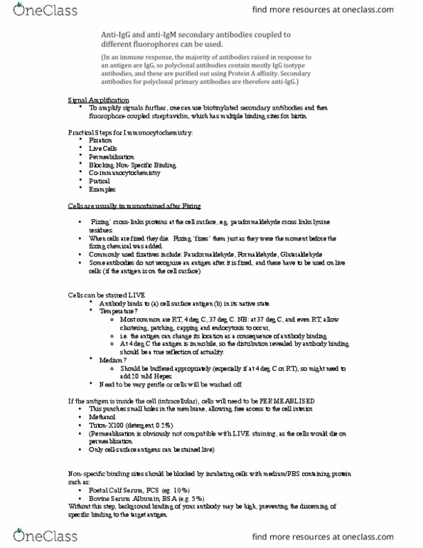
The most versatile way of doing fluorescence immunostaining is to use fluorescently conjugated
species specific secondary antibodies that recognise the –FC portion of the primary antibody rather
than directly coupling each primary antibody to a fluorophore.
Thus it is important to note the species in which an antibody was raised.
•
•
o
•
o
•
•
•
•
•

Signal Amplification
To amplify signals further, one can use biotinylated secondary antibodies and then
fluorophore-coupled streptavidin, which has multiple binding sites for biotin.
Practical Steps for Immunocytochemistry:
Fixation
Live Cells
Permeabilisation
Blocking Non-Specific Binding
Co-immunocytochemistry
Pratical
Examples
Cells are usually immunostained after Fixing
‘Fixing’ cross-links proteins at the cell surface, e.g. paraformaldehyde cross links lysine
residues. !!!
When cells are fixed they die. Fixing ‘fixes’ them just as they were the moment before the
fixing chemical was added.
Commonly used fixatives include: Paraformaldehyde, Formaldehyde, Glutaraldehyde
Some antibodies do not recognise an antigen after it is fixed, and these have to be used on live
cells (if the antigen is on the cell surface)
Cells can be stained LIVE
Antibody binds to (a) cell surface antigen (b) in its native state.
Temperature?
Most common are RT, 4 deg C, 37 deg C. NB: at 37 deg C, and even RT, allow
clustering, patching, capping and endocytosis to occur,
i.e. the antigen can change its location as a consequence of antibody binding
At 4 deg C the antigen is immobile, so the distribution revealed by antibody binding
should be a true reflection of actuality.
Medium? !
Should be buffered appropriately (especially if at 4 deg C or RT), so might need to
add 20 mM Hepes.
Need to be very gentle or cells will be washed off.
If the antigen is inside the cell (intracellular), cells will need to be PERMEABLISED
This punches small holes in the membrane, allowing free access to the cell interior
Methanol
Triton-X100 (detergent, 0.2%)
(Permeablisation is obviously not compatible with LIVE staining, as the cells would die on
permeablisation.
Only cell-surface antigens can be stained live)
Non-specific binding sites should be blocked by incubating cells with medium/PBS containing protein
such as:
Foetal Calf Serum, FCS (eg. 10%)
Bovine Serum Albumin, BSA (e.g. 5%)
Without this step, background binding of your antibody may be high, preventing the discerning of
specific binding to the target antigen.
•
•
•
•
•
•
•
•
•
•
•
•
•
•
o
o
o
•
o
•
•
•
•
•
•
•
•
Document Summary
Binding to a foreign target causes that b-lymphocyte to divide to produce a large clonal population of cells that each produce lots of the target-recognising antibody. We can exploit this to produce antibodies that specifically recognise a part of a protein that we want to be able to bind. The antibodies that recognise a target antigen of interest e. g. gap43 or actin, in the examples given are known as primary antibodies. Primary antibodies can be raised in different species, commonly mice, rabbits, goats or sheep. Two classes of primary antibodies used: o o. These are derived from a single b-cell clone, and all are identical and recognise a single specific site on the antigen. Usually raised in mice e. g. mouse antihuman actin monoclonal antibody. These are derived from several b-cell clones, and are a mixture of antibodies that all recognise the same antigen but different target sites on it.