HTHSCI 1DT3 Lecture Notes - Lecture 7: Die Shrink, Centrosome, Endocytosis

23 Jun 2018
School
Department
Course
Professor

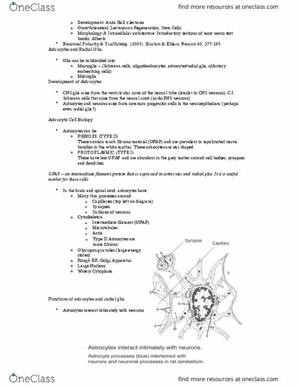
Astrocytes and Radial Glia
These are cells found in the central nervous system only
Astrocytes are stellate cells (star shaped), while radial glia are elongated in
morphology.
Radial glias are like astrocytes, but have a particular shape. They provide a track
for neuronal cells to migrate along.
Number of Cells:
In the human cerebral cortex there are over 10 billion neurons
Ratio of neurons to glia is typically 2:1
However, taken as a whole, in the brain there are probably more glia than
neurons as oligodendrocytes are abundant in white matter.
White matter has greater oligodendrocytes (support cells) than neurons (vs. grey
matter).
In addition to the major classes of nervous system cells, there are also:
Endothelial Cells: of the nervous system vasculature, essential for forming the
blood-brain barrier
Fibroblasts: in the meninges (membranes surrounding the brain, under the
scalp) and
Ependymal cells: (lining the ventricles)
The ventricles and central canal running down the central core of the spinal cord are
filled with cerebrospinal fluid (CSF), secreted by the ciliated ependymal cells that line
them.
•
•
•
•
•
•
•
•
•
•

Cells 2: Neurons and Astrocytes – Morphology, Function and Cell Biology
8th October 2012
Peripheral Nervous System vs. Central Nervous System Neurons
Neurons that have their cell bodies outside the brain or spinal cord are called peripheral
neurons. The peripheral nervous system are neurons outside the brain and spinal cord.
Those that have their cell bodies inside the brain or spinal cord are called central neurons. The
central nervous system covers are inside the brain and spinal cord.
N.B. It is the location of the CELL BODIES that determine whether a neuron is CNS or PNS. E.g. motor
neurons extend through the periphery, but are still CNS neurons as their cell bodies are found in the
spinal cord.
Function of the Neurons: To receive electrical and chemical signals from other neurons and transmit
signals to other neurons in the form of action potentials and released neurotransmitters.
Specific functions: depend on the neuron in question, e.g.:
Interneurons: are CNS neurons that communicate with other CNS neurons
Motor Neurons: innervate muscle and stimulate muscle contraction
Sensory Neurons: (e.g. in the skin, ear and tongue) transmit information about the surrounding
environment to central nervous system neurons.
These transmit signals from outside the CNS to inside but have cell bodies outside the CNS (e.g.
Dorsal Root Ganglion)
Neuronal Development
CNS Neurons arise from the neural tube (the neural tube later becomes the central canal and
ventricles)
PNS neurons arise from the neural crest (these neural crest cells surround the neural tube)
The neural tube develops into the ventricles, filled with cerebrospinal fluid
The neural tube is lined with a specialized epithelium known as neuroepithelium, and these cells
divide to form the brain and spinal cord, giving rise to all CNS neurons (and astrocytes).
The dividing neuroepithelial cells (stem cells/neuroblasts) have an elongated morphology and
span the whole width of the neural tube (i.e. from the ventral surface to the dorsal surface –
NOT cranio/caudally) - (contacting the pial/outer surface and the ventricular/inner surface).
i.e. outer surface of brain, to inner surface of ventricle lining.
The cell body of the neurons translocates away from the ventricular surface (as shown on the
left side of the diagram below), then returns, divides and re-contacts the pial surface.
Neurons divide in (near) the ventricular zone only (right side of diagram below)
•
•
•
•
•
•
•
•
•
•
•
••

Once generated, cortical neurons move away from the ventricular zone towards the outer
surface to lodge in the cortical plate. The cortical plate is the outer regions of the brain (i.e.
cortex).
The cortical plate develops from inside out, and neurons use the radial glia as a guide (long
elongated pinkish-red cells spanning ventricular zone to marginal zone). The earliest generated
neurons (lighter green) reach first, and as the cortical plate develops, newer neurons (darker
green) pass the earlier ones and develop the external layers ‘higher up’ in the brain.
The cortical plate refers to the outer regions of the brain (i.e. cortex), and earlier on in
development, the neurons in light green migrate up using the radial glia as a guide. As
development progresses, newer neurons (dark green) migrate up past the older neurons (light
green), and are positioned further outwards.
•
•
•