KINESIOL 1Y03 Lecture Notes - Lecture 16: Ulnar Nerve, Central Canal, Lumbar Vertebrae

20 Apr 2016
School
Department
Course
Professor

Document Summary
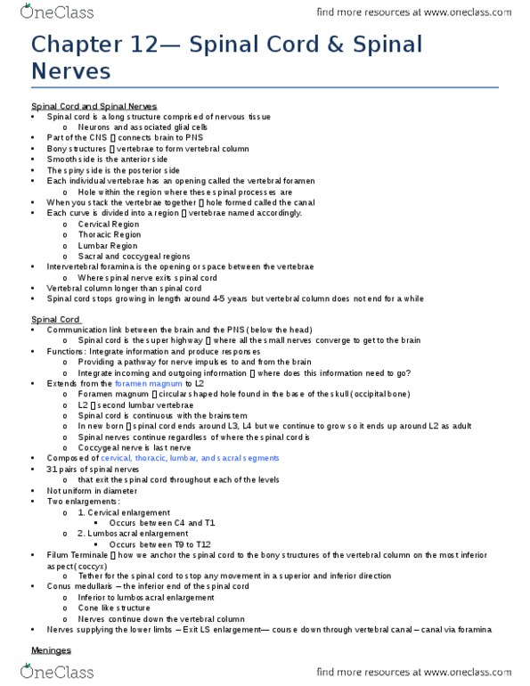
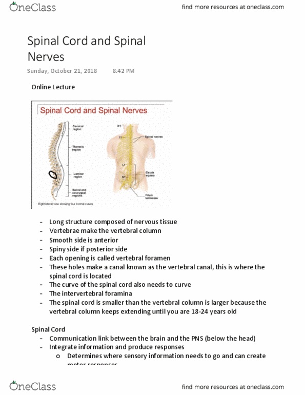
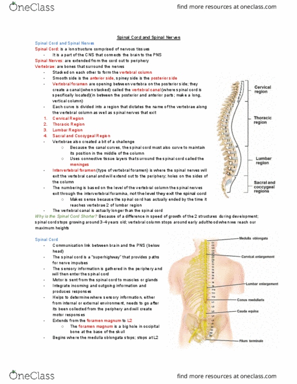
The spinal cord is the long structure comprised of nervous tissue. Its part of the cns and connects the brain to the pns. Made of nervous tissue which is neurons and associated glial cells. On the left side of the diagram, there is bony structure surrounding the cord called vertebrae and are stacked onto one another to form the vertebrae column. The smooth side of the vertebrae is the anterior side and spiny process is the posterior side. In the region where you see these spiny processes, each individual vertebrae has an opening called the vertebral foramen. That"s a hole within the region of where these spiny processes are. When you stack the vertebrae on top of each other, the successive holes then creates a canal, called the vertebral canal, and this is where the spinal cord itself is located. The vertebral column have several curves in it and each is divided into several regions.