MEDRADSC 3C03 Lecture Notes - Lecture 7: Acute Liver Failure, Fresh Frozen Plasma, Liver Biopsy

8 Jun 2020
School
Department
Course
Professor
Document Summary
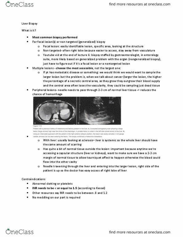
For focal lesion(s) or non targeted (generalized) biopsy. Biopsies are used to diagnose liver disease & measure extent of the damage ie) hcc vs staging of hepatitis. The liver is of the most common organs for appearance of metastasis. L lobe: immediately posterior is the ivc & abdominal aorta, stomach, right portal vein & bile ducts, duodenum anterior, proximity to the pancreas. R lobe: safer & wider area without vasculature or other organs - have to be aware of the gallbladder though. Peripheral lesions: needle should pass through 2-3 cm of normal liver tissue - reduces the chance of bleeding/hemorrhage. Not the safest, largest may be necrotic at the center. Us contrast enhanced micro-bubble imaging is used more frequently to diagnose the etiology of the liver lesions, may reduce need for core biopsies - diagnose type of cancer. Lack of knowledge on the type of imaging needs to be done for c+ imaging to be used more.