PHGY 214 Lecture Notes - Lecture 25: Qrs Complex, Electric Current, Qt Interval

8 Dec 2017
School
Department
Course
Professor
Document Summary
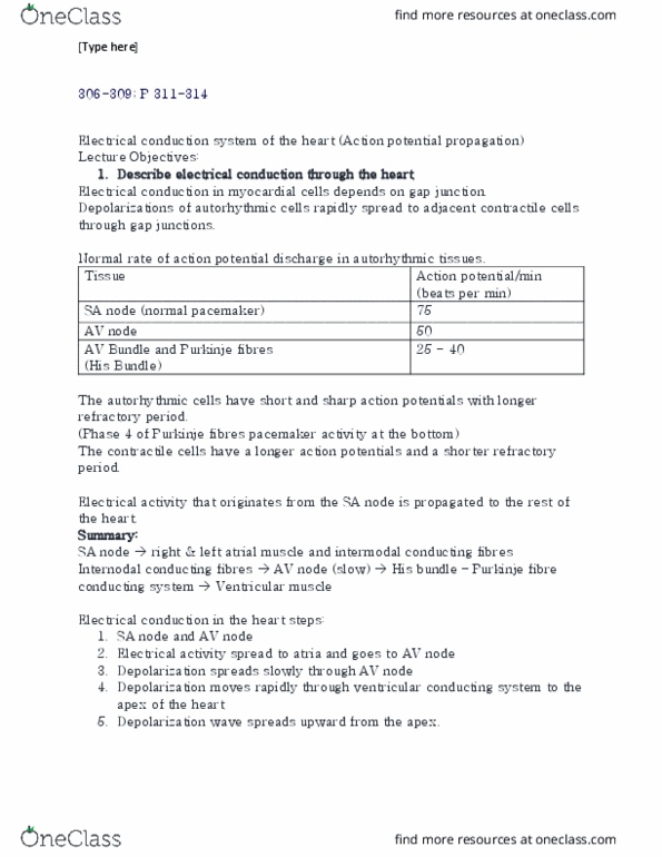
Sa node internodal pathways: anterior, middle and posterior. Sa and av node have pacemaker potential electrical activity spread. Sa node depol atrial muscle (l or r= bockmans bundle) or intermodal conducting fibers av node (slow) bundle of his and purkinje conducting sys ventricular sys rapid depol spreads up from apex. 1st layer is endocardium and last layer depol is epicardium. Voltage diff b/n excited and non-excited parts of myocardium. Ecg is a recording of electrical activity of the heart from body surface: 12 ecg leads: 6 chest leads: v1 to v6 unipolar (only use one electrode: give representation on horizontal plane, use angle of lewis to det where to put the leads. At rest, no voltage diff outside of the cell (flat line) Depol waves detected if wave is travelling twrds +ve electrode= increasing deflection. After depol there is no volt diff outside cell memb so flat line.