PHGY 214 Lecture Notes - Lecture 27: Blood Proteins, Heart Block, Myopathy

8 Dec 2017
School
Department
Course
Professor
Document Summary
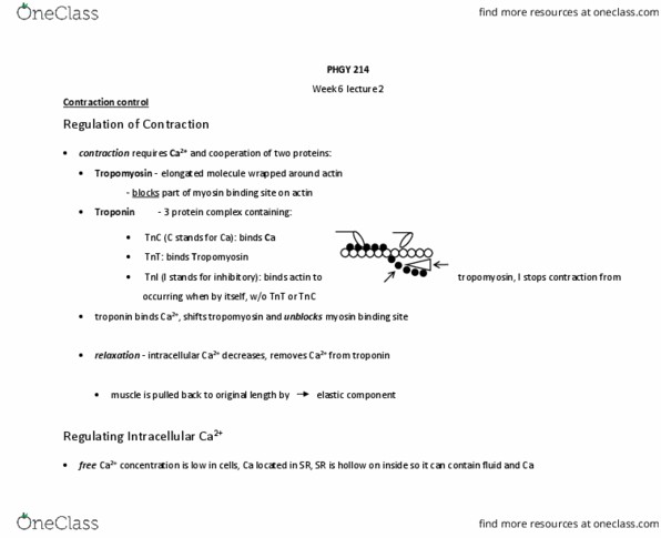
Excitation-contraction coupling (ecc) larger t tubule allows for ap w/o stim intercalated discs allow for rapid spread of ap structure: actin twirls into double helix form, troponin: binds to tropomyosin, actin and. Ecc: ap in contractile cell travels down t-tubule, ap enters from adjacent cell (other cardiac) connected via gap junctions, ap gen leads to v. g. In skeletal muscle: voltage induced ca release instead: dihydropyridine rs: open ryr rs (called this b/c can be blocked by hydropyridine molecule) Ne and e hormones released form adrenal medulla into blood= fight or flight. Ve inotropic (contraction) effect: +ve dromotopoc (conduction velocity) effect, +ve chronotropic (heart rate) effect, +ve lusitropic (relation) effect. L-type ca chan, ryr (ca release chan) and serca (transfers ca from cytosol to lumen of sr) increase in activity if serca is more active it pulls ca off troponin complexes causing relaxation. Can be graded by how much ca enters the cell. Sa node depol atrial muscle (l and dispersion.