HK 3401 Lecture Notes - Lecture 16: Pericardial Sinus, Coronary Sinus, Pulmonary Artery

11 Nov 2016
School
Department
Course
Professor

Document Summary
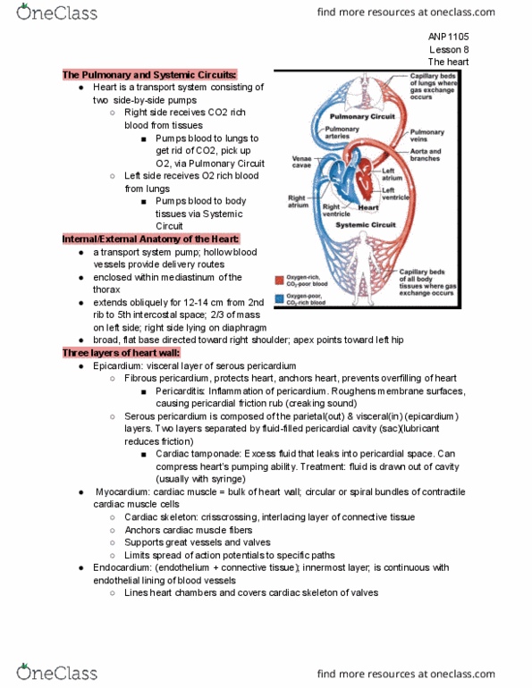
The heart is a double self-adjusting muscular pump, the parts of which work in unison to propel blood to the body: the right side of the heart receives poorly oxygenated blood from the body through the. Faces posteriorly towards the bodies of vertebrae t6-t9 and is separated from them by the pericardium, oblique pericardial sinus, esophagus and aorta. Extends superiorly to the bifurcation of the pulmonary trunk and inferiorly to the coronary sulcus (groove). Receives pulmonary veins on the right and left sides of the left. !1 atrium and the superior and inferior vena cava at the superior and inferior ends of the right. Wednesday, november 2, 2016 atrium: four surfaces of the heart: Anterior (sternocostal) surface: formed mainly by the right ventricle. Diaphragmatic (inferior) surface: mainly by left ventricle and partly by right ventricle; it is related to the central tendon of the diaphragm.