MCB 2050 Lecture Notes - Lecture 4: Fluorescence Microscope, Hybridization Probe, Contig

8 Sep 2018
School
Department
Course
Professor
Document Summary
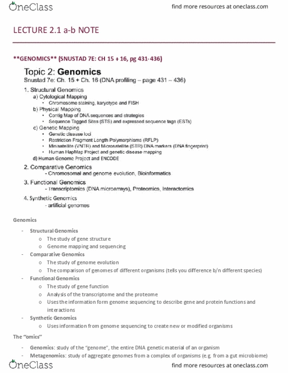
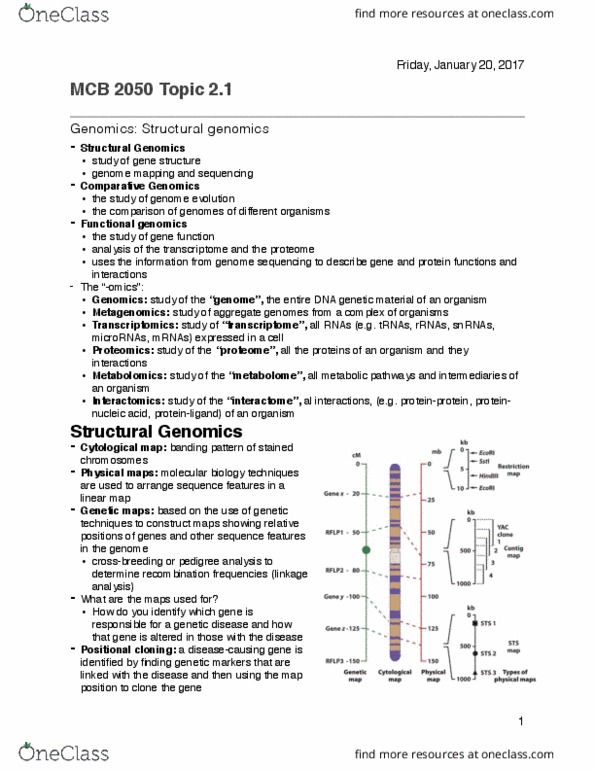
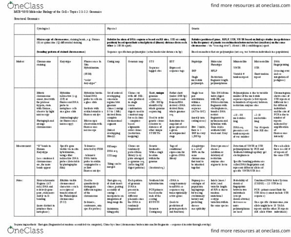
Mcb*2050 molecular biology of the cell topics 2. 1-2. 2: genomics. Genetic microscopy of chromosome, staining bands, e. g. giemsa (g) or quinacrine (q) differential staining (banding pattern of stained chromosomes) Mitotic chromosome smear, treat with the protease trypsin, stain with giemsa, observe by light microscopy. Hybridize radioactive (e. g. 32p) or fluorescent dna probe to metaphase cells on a glass slide. Specific gene visible via in situ hybridization, using either radioactive dna probes (detectable by photographic film) Hybridize biotin- bound dna probe to cells on a glass slide. Incubate with avidin bound to a fluorescent dye. Telomeric dna- biotin probe to avidin conjugated to a yellow fluorescent dye set of ordered overlapping genomic clones (contigs) 0. 5 to. 1 mb each set of ordered contigs for each chromosome. The comparison of genomes of different organisms. Analysis of the transcriptome and the proteome. Uses the information from genome sequencing to describe gene and protein functions and interactions.