PSYC 4600 Lecture Notes - Lecture 6: Visual Cortex, Optic Chiasm, Right Fielder

15 Nov 2016
School
Department
Course
Professor
Document Summary
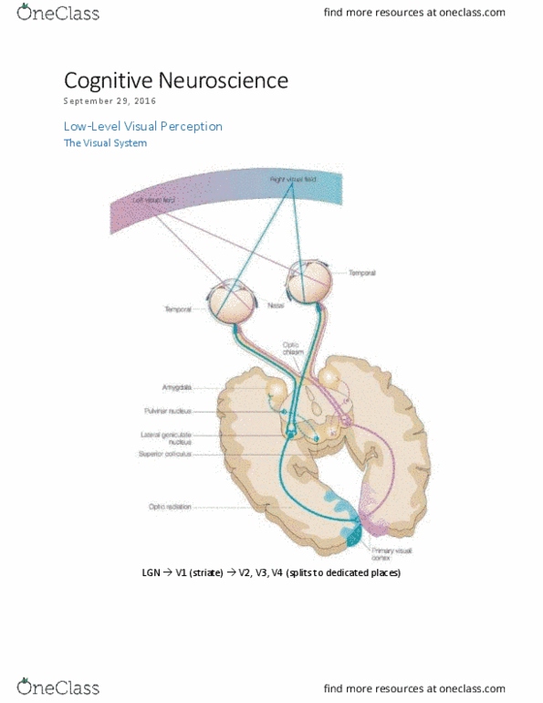
Lgn -> v1 (striate) -> v2, v3, v4 (splits to dedicated places) Retinotopic topography: the way the brain is organized an how that corresponds to how info is processed. What areas correspond to what processing: organized spatially. 2 regions close together will project to neurons that are close together in the visual cortex. Watching flashing bullseye pattern -> injected with radioactive glucose to show neural activity (uptake form active neuron) so it could be exposed to radioactively sensitive film to act as a marker. Right visual field -> left hemisphere of striate. Areas are spatially relevant (in the same kind of pattern/map: cortical magnification. Info processed by the fovea (f) in the center, there is more processing of that information. Information in your direct line of focus is processed the most. Superior (s) visual field projects to inferior (i) cortex and vice versa. We know the image is switched in the back of the eye.