ZOO 3200 Lecture 4: Membrane Transporters

2 May 2018
School
Department
Course
Professor

Types of membrane transport and transporters
•
Discovery of Aquaporins
•
Diversity of ion channels involved in passive diffusion
•
Active transport
•
Outline:
Readings: Ch.5 (pages 99-124, 439,440; Fig 16.16) & Discovery of
Aquaporins
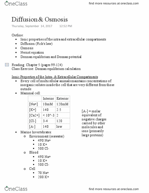
Every cell maintains concentrations of inorganic solutes
inside the cell that are very different from those outside
•
[Na+] -higher outside
○
[K+] -higher inside
○
[Ca2+] -higher outside
○
[Cl-] -higher outside
○
[A-] -higher inside
○
Need to know how the concentrations vary from the interior
to the exterior:
•
Ionic Properties of the Intra-& Extracellular Compartments
Simple Diffusion (e.g. O2, CO2, hydrophonic solute)
a.
By channel protein (e.g. ions, water)
!
By carrier protein (e.g. urea, glucose)
!
Facilitated Diffusion
b.
Passive -requires no energy; direction of solute movement is
from high to low concentrations
1.
Active -requires ATP; direction of solute movement is from
low to high concentrations (e.g. H+, Na+)
2.
Types of Membrane Transport
The saturation kinetics of facilitated transporters follows a
hyperbolic relationship (e.g. Michaelia-Menton kinetics)
•
*see figure on slide
•
Simple diffusion has a slow rate of uptake
○
Facilitated diffusion reaches a maximum rate (of glucose
uptake) at Vmax
•
[S] that gives 1/2 Vmax = Km (Michaelis constant)
•
Michaelis-Menton equation: V = (Vmax*[S]) / ([S] + Km)
•
Kinetics of Facilitated Transport
E.g. glucose and Na+ channel
○
Uniporter
•
E.g. Na+/K+ ATPase and Cl-/HCO3-exchanger
○
Antiporter
•
E.g. NKCC = Na+, K+, 2Cl-cotransporter and K+/Cl-
cotransporter
○
Symphorter
•
Types of Transporters
Determines DNA and amino acid sequences of the
water channel AQP1
○
Used Xenopus eggs and artificial cells to demonstrate
that AQP1 is responsible for osmosis
○
Determined the 3D structure of AQP1 and how the
water channel works
○
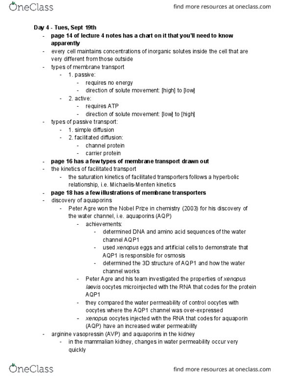
Peter Agre won the Nobel Prize in Chemistry (2003) for his
discovery of the water channel (i.e. aquaporins or AQP)
•
They compared the water permeability of control
oocytes where the AQP1 channel was over-expressed
○
They found that the oocytes injected with the RNA that
codes for AQP have an increased water permeability
(the volume of the cell would increase until it ruptured)
○
Peter Agre and his team investigated the properties of
Xenopus laevis oocytes microinjected with the RNA that
codes for the protein AQP1
•
In the mammalian kidney, changes in water
permeability occur very quickly
○
AVP is a hormone that regulates water permeability
○
*see figure
○
This activates protein kinase A which causes
storage vesicles containing AQP2 to fuse with
the membrane of the collecting duct, allowing
the water to move into the collecting duct cell
and then from the collecting duct cell (via
AQP3) into the peritubular capillary
!
AVP moves down the peritubular capillary, and into
the extracellular fluid where it binds to a vasopressin
receptor causing the release of cAMP
○
Arginine Vasopressin (AVP) and Aquaporins in the Kidney:
•
Discovery of Aquaporins
Voltage-gated channels open/close in response to changes in
membrane potential
a.
Ligand-gated channels open/close in response to
presence/absence of ligand
b.
Mechanically-gated channels open/close in response to
changes in cell shape
c.
Diversity of Ion Channels involved in Facilitated Diffusion
Maintaining high [K+] in and [Na+] out
!
Maintaining the transmembrane electrical
potential
!
Most common is the Na+-K+ pump responsible for:
○
The protein hydrolyzes ATP to obtain
ATP-bond energy to carry out active ion
transport
□
The proton pump (H+/K+ ATPase) that secretes
stomach acid is a protein that is expressed in
parietal cells
!
The pump exchanges two H+ for two K+ during
each pumping cycle (it is therefore
electroneutral)
!
The proton-pump protein molecules are
positioned in the portions of the apical
membrane that line the canaliculi
□
The apical membrane of each parietal cell
projects into the cells as invaginations called
canaliculi (increases SA)
!
Electroneutral active transport is responsible for
secretion of stomach acid in the vertebrae stomach
lining
○
Primary -the energy released by ATP hydrolysis drives
solute movement against an electrochemical gradient
•
Na+/K+ ATPase: moves 3 Na+ out of the cell
and 2 K+ into the cell to produce ATP (against
the electrochemical gradient)
!
Na+/glucose cotransporter: transports glucose
and 2 Na+ into the cell (in the direction of the
electrochemical gradient)
!
An example is the active transport of glucose in the
small intestine
○
Secondary -the movement of an ion down its
electrochemical gradient provides the energy to drive
cotransport of a second solute against its electrochemical
gradient
•
Active Transport
*see figure
•
Glut2 and Glut5 are uniporters
•
SGLT1 is the Na+/glucose symporter
•
The facilitated diffusion transporter (with one solute) moves
against the electrochemical gradient
•
ATPase and cotransporter diffuse with the electrochemical
gradient
•
Types of Monosaccharide Transporters
Membrane Transporters
Tuesday,+ September+ 19,+2017
1:01+PM
Document Summary
Diversity of ion channels involved in passive diffusion. Readings: ch. 5 (pages 99-124, 439,440; fig 16. 16) & discovery of. Ionic properties of the intra- & extracellular compartments. Every cell maintains concentrations of inorganic solutes inside the cell that are very different from those outside. Need to know how the concentrations vary from the interior to the exterior: Passive - requires no energy; direction of solute movement is from high to low concentrations a. b. Active - requires atp; direction of solute movement is from low to high concentrations (e. g. h+, na+) The saturation kinetics of facilitated transporters follows a hyperbolic relationship (e. g. michaelia-menton kinetics) Facilitated diffusion reaches a maximum rate (of glucose uptake) at vmax uptake) at vmax. Simple diffusion has a slow rate of uptake. [s] that gives 1/2 vmax = km (michaelis constant) Michaelis-menton equation: v = (vmax*[s]) / ([s] + km) Nkcc = na+, k+, 2cl- cotransporter and k+/cl- cotransporter.