ANP 1105 Lecture 4: Anatomy & Physiology ANP1105 section 4.2 heart notes

9 Dec 2016
School
Department
Course
Professor
Document Summary
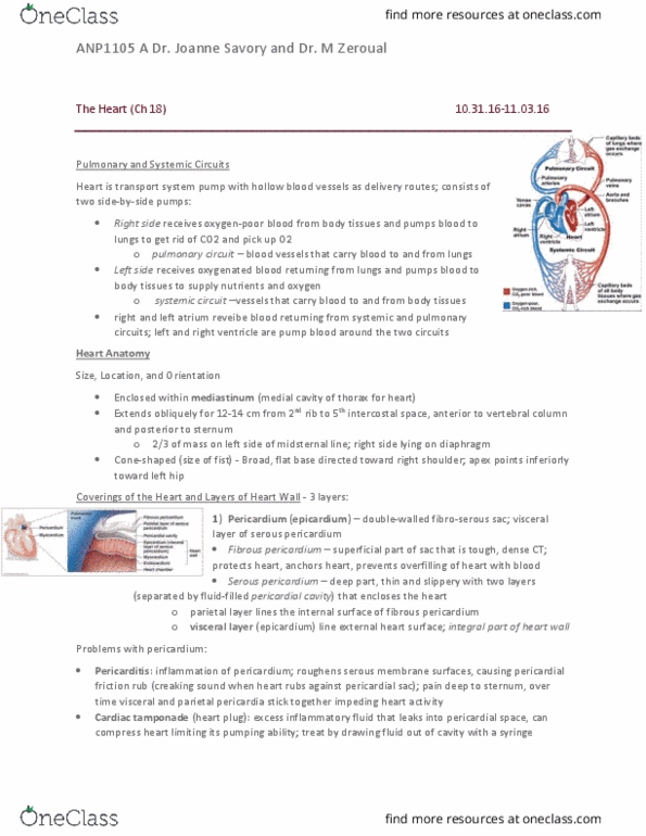
Location; superior surface of diaphragm, left of the midline, from 2nd rib to 5th intercostal space; a(cid:374)terior to the verte(cid:271)ral (cid:272)olu(cid:373)(cid:374), posterior to the ster(cid:374)u(cid:373); (cid:862)(cid:373)ediasti(cid:374)u(cid:373)(cid:863). The three layers of the heart is: Peri(cid:272)ardiu(cid:373)" a dou(cid:271)le-walled sac around the heart composed of; The parietal layer lines of the internal surface of the fibrous pericardium. The visceral layer or epicardium lines the surfaces of the heart. They are separated by the fluid-filled pericardial cavity. Prevents overfilling of the heart with blood. Allows for the heart to work in a relatively friction-free environment. Epicardium visceral layer of the serous pericardium. * exam will have diagram of heart either anterior or asterior another question is understanding the difference between arteries and veins, veins deliver blood to the heart, arteries deliver the blood away. Myocardium cardiac muscle layer forming the bulk of the heart. Provides support for great vessels and valves. Directs spread and action potentials across the heart.